
Fig. 10
- ID
- ZDB-FIG-120202-50
- Publication
- Dohn et al., 2012 - Distinct phases of Wnt/β-catenin signaling direct cardiomyocyte formation in zebrafish
- Other Figures
- All Figure Page
- Back to All Figure Page
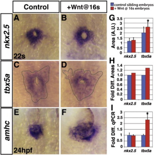
Effects of increased Wnt signaling during atrial CM differentiation are evident by the 22 somite stage. (A,C,E) HCSEs. (B,D,F) GFP + embryos with increased Wnt signaling at the 16 s stage. (B) The amount of cells expressing nkx2.5, which at 22 s primarily marks ventricular cells, is not increased when Wnt signaling is increased during cardiac differentiation. (D) The amount of cells expressing tbx5a, which is expressed in both ventricular and atrial cells, is modestly increased by 22 s. (F) When Wnt signaling is increased at 16 s, amhc expression indicates that the atria are wider compared to controls (E) by 24 hpf. (G) Areas of the amount of cells expressing nkx2.5 and tbx5a in arbitrary units. (H) Fold difference in the areas of cells expressing nkx2.5 and tbx5a. (I) qPCR analysis of nkx2.5 and tbx5a expression at 22 s. There is an increase in tbx5a greater than what is observed with the ISH analysis. However, we did observe an increase in tbx5a is expressed in the forelimb mesenchyme (data not shown). Therefore, the increase in tbx5a expression observed with qPCR likely reflects an increase in tbx5a expression from both populations. No difference in tbx5a expression in the eye was observed between HCSEs and embryos with increased Wnt signaling at the 16 s stage at the 22 s stage (data not shown). Asterisks indicates significant difference using Student′s t-test. |
Reprinted from Developmental Biology, 361(2), Dohn, T.E., and Waxman, J.S., Distinct phases of Wnt/β-catenin signaling direct cardiomyocyte formation in zebrafish, 364-76, Copyright (2012) with permission from Elsevier. Full text @ Dev. Biol.