
Fig. 8
- ID
- ZDB-FIG-120202-48
- Publication
- Dohn et al., 2012 - Distinct phases of Wnt/β-catenin signaling direct cardiomyocyte formation in zebrafish
- Other Figures
- All Figure Page
- Back to All Figure Page
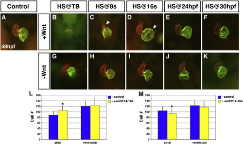
Wnt signaling is necessary and sufficient to promote atrial cell development during CM differentiation. (A) Representative HCSE. (B–F) GFP + embryos with increased Wnt signaling. (G–K) GFP + embryos with decreased Wnt signaling. Decreasing Wnt signaling from TB through 30 hpf had inconsistent and modest effects on morphology. (B,C) Increasing Wnt signaling at TB or 8 s almost eliminates and greatly reduces the size of the hearts, respectively, at 48 hpf. Arrow in C indicates the severely smaller ventricle. (D) When Wnt signaling is increased at 16 s, the atrium is significantly enlarged (arrow). Although the ventricles were dismorphic, we did not find an effect on ventricular cell number. (E) Increasing Wnt signaling at 24 hpf causes modestly enlarged atria. (F) Increasing Wnt signaling at 30 hpf no longer has a discernible effect on the heart. (L) Increasing Wnt signaling at 16 s causes a specific increase in atrial cell number. (M) Decreasing Wnt signaling at 16 s causes a modest reduction in atrial cell number. Images are frontal views. Red indicates ventricle and green indicates atrium. Asterisks indicate a significant difference in the cell number from HCSEs and GFP + embryos in Supplemental Table 3 according to Student′s t-test. |
Reprinted from Developmental Biology, 361(2), Dohn, T.E., and Waxman, J.S., Distinct phases of Wnt/β-catenin signaling direct cardiomyocyte formation in zebrafish, 364-76, Copyright (2012) with permission from Elsevier. Full text @ Dev. Biol.