|
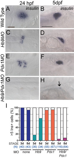
ins mRNA expression in hb9 and pdx1 morphants. In situ detection of ins mRNA in wild-type embryos (A,B), hb9 morphants (C,D), pdx1 morphants (E,F) and hb9/pdx1 double morphants (G,H) at 24 h post fertilization (hpf) and 5 days post fertilization (dpf). In hb9 and pdx1 morphants the number of ins expressing cells is strongly reduced at 24 hpf as compared to control embryos (A,C,E) but has substantially increased by 5 dpf (D,F). (G,H) ins expression is missing in most double morphants (arrow in H marks a single ins positive cell). Embryos are shown from ventral (24 hpf) and lateral (5 dpf) view with anterior to the right. Scale bars correspond to 20 μm (24 hpf) and 50 μm (5 dpf). (I) Proportion of embryos showing > 5 Ins+ cells in control and morphant embryos at 3 dpf and 5 dpf. Beta cells emerge between 3 dpf and 5 dpf in hb9 and pdx1 morphants, while there is persistent absence in the hb9/pdx1 double morphant.
|