
Fig. S9
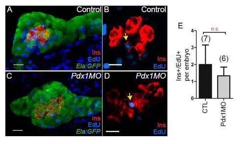
Proliferation during exocrine pancreas formation. Tg(ela3l:EGFP)gz2 [35] embryos labeled with EdU from 24 to 72 h post fertilization (hpf), and fixed at 84 hpf. EdU detection (blue) was followed by antibody staining to label Elastase (Ela)-expressing exocrine cells (Ela:green fluorescent protein (GFP), green) and beta cells (insulin (Ins), red). (A,C) Three-dimensional confocal projections showing composite of Ela:GFP, EdU and Ins in control (A), and pdx1 morphant embryos (C) at 84 hpf. EdU labels exocrine pancreas extensively in control and morphants. (B,D) Single plane views of embryos as in (A) and (C), showing rare cells with EdU/Ins colabeling (arrow). For clarity, the GFP channel is not shown. (E) Quantitation of EdU/Ins colabeled cells per embryo in control and pdx1 morphant embryos, showing mean and standard deviation. NS, not significant; P > 0.05 as determined by unpaired t test. Anterior is to the left. Scale bar = 15 μm. |