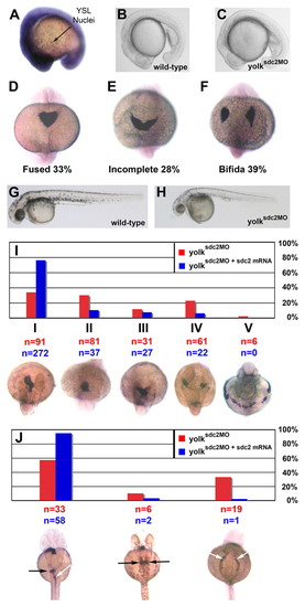
Impaired cardiac primordia and primitive foregut migration in yolksdc2MO embryos. (A) Ubiquitous expression of sdc2 at the 16-somite stage. Strong expression is seen throughout embryonic tissues and within individual yolk syncytial nuclei. (B,C) Lateral views of wild-type (B) and yolksdc2MO (C) embryos at the 18-somite stage. Patterning of yolksdc2MO embryos is relatively normal at this stage. (D-F) Expression of cmlc2 in embryos at the 22-somite stage (dorsal view). Bilateral cardiac primordia were completely fused in 100% of wild-type embryos but only in 33% of yolksdc2MO embryos (D) at this stage. Cardiac primordia migration was defective in yolksdc2MO embryos, with 28% manifesting incomplete heart-field fusion (E) and 39% with cardia bifida (F). (G,H). Lateral views of wild-type (G) and yolksdc2MO (H) embryos at 40 hpf. The overall patterning in yolksdc2MO embryos remains normal, but the embryos are smaller, with a defective posterior yolk tube, larger yolk cell, pericardial edema and decreased pigmentation (H). (I) Bar graph depicting the spectrum of cardiac defects seen in yolksdc2MO embryos (red bars) and yolksdc2MO embryos co-injected with sdc2 mRNA (blue bars) at 40 hpf. Below the graph, the expression of cmlc2 is shown in embryos displaying the spectrum of cardiac phenotypes seen in yolksdc2MO embryos (dorsal view, anterior up for class I-IV embryos; ventral view, anterior up for class V embryo). Cardiac phenotypes vary in severity from left to right and include normal heart tube looping (class I), impaired elongation of the cardiac cone to form the primitive heart tube (class II), incomplete fusion of bilateral heart fields (class III), cardia bifida (class IV) and a band-like distribution of myocardial cell precursors (class V). Numbers of embryos displaying each phenotype are noted in red for yolksdc2MO embryos and in blue for yolksdc2MO + sdc2 mRNA embryos. In rescued embryos, the percentage of embryos with cardiac anomalies decreased from 66% to 24% (P<0.01). (J) Bar graph depicting the spectrum of gut defects seen in yolksdc2MO embryos (red bars) and yolksdc2MO embryos co-injected with sdc2 mRNA (blue bars) at 40 hpf. Below the graph, the expression of foxa3 is shown in embryos displaying the spectrum of gut phenotypes seen in yolksdc2MO embryos (dorsal view, anterior up). From left to right, embryos display normal gut looping (white arrow) and asymmetric hepatic/pancreatic bud formation (black arrow), duplicated hepatic/pancreatic buds (black arrows), and bilateral condensation of the posterior endoderm resulting in the formation of a two-sided foregut (white arrows).
|
