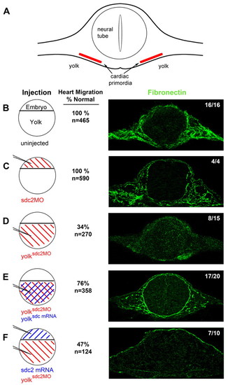
Extra-embryonic Sdc2, but not embryonic Sdc2, is required for fibronectin fibrillogenesis throughout the embryo. (A) Diagram of an embryo cut in cross-section at the level of the cardiac primordia. Fibronectin fibrils form in the extracellular space adjacent to the cardiac primordia and surrounding the neural tube. (B-F) Transverse sections (right) of 22-somite embryos immunostained for fibronectin (green). Circular diagrams (left) represent different injection patterns of sdc2 morpholino (red) and sdc2 mRNA (blue). (B) Uninjected wild-type embryos (16/16 embryos) and (C) traditional sdc2 morphants (4/4 embryos) had normal fibrillogenesis, indicating that embryonic Sdc2 is not required for fibrillogenesis. (D) yolksdc2MO morphants had no fibril formation in 8/15 embryos. With co-injection of sdc2 mRNA into the yolk at the dome stage, fibrillogenesis was rescued in 17/20 embryos (E). By contrast, injection of sdc2 mRNA at the 1-to-2-cell stage, to overexpress Sdc2 in embryonic cells, followed by injection of sdc2 morpholino into yolk at the dome stage (F), did not rescue fibrillogenesis (7/10 embryos). As noted by the numbers adjacent to the confocal microscopy images, a higher percentage of normal cardiac migration correlates with the presence of fibronectin fibrils.
|
