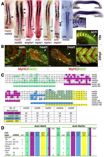
Mef2d and Mef2c expression during zebrafish skeletal myogenesis and cardiogenesis. (A) In situ mRNA hybridisation of mef2d, mef2c, myod, actc1 (actc), smyhc1 and myhz1 viewed in dorsal (anterior to top) or lateral (right-most panels; anterior to left, dorsal up) flatmount. Mef2d, myod and actc1 mRNAs coincide in early adaxial slow (black arrow) and lateral fast (black arrowheads) myoblasts and precede smyhc1 and myhz1 in differentiated slow (bracket) and fast muscle fibres. Sequential expression of actc1 and myhz1 during fast muscle differentiation is confirmed in smu (smo) mutant, which lacks slow muscle. Mef2c expression commences as adaxial cells form slow fibres (red arrow) and in bilateral heart fields (green arrowhead). Mef2c expression in fast muscle parallels terminal differentiation. TB, tailbud; s, somite. (B) Immunodetection of Mef2 and MyHC in wholemount embryos viewed in dorsal (heart; anterior to top) or lateral [somite (som); anterior to left, dorsal up] flatmount. Confocal single scans of anti-Mef2c and z-stacks of anti-Mef2 antibodies detect muscle nuclei in skeletal myoblasts in the presomitic mesoderm (arrow), muscle fibres, heart tube and undifferentiated cardiomyoblasts (arrowheads). Scale bars: 20 µm. (C) Sequence alignment of start codon region (blue arrow) of five zebrafish Mef2 genes and comparison with MOs employed. Coloured squares highlight identity with the MO. The table shows the number of mismatches. (D) Location of anti-Mef2 and anti-Mef2c immunoreactivity in relation to mef2d and mef2c mRNAs and the effect of the indicated MO combinations (see Figs S1-S3 in the supplementary material). +, ± and-indicate normal, low and no nuclear reactivity, respectively; where there is no entry, nuclear reactivity was not determined.
|
