FIGURE

Fig. 1

ID
ZDB-FIG-061226-15
Publication
Shimizu et al., 2006 - Cdx-Hox code controls competence for responding to Fgfs and retinoic acid in zebrafish neural tissue
Other Figures
All Figure Page
Back to All Figure Page
Fig. 1

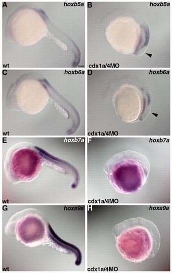
Loss of posterior hox expression in embryos lacking Cdx1a/4. Expression of hoxb5a (A,B), hoxb6a (C,D), hoxb7a (E,F) or hoxa9a (G,H) in wild-type controls (wt) (A,C,E,G) and in embryos that received an injection of 1 ng cdx1aMO and 1 ng cdx4MO (B,D,F,H) at 22 hpf. hoxb5a, hoxb6a, hoxb7a and hoxa9a are expressed in the spinal cord. hoxb5a is expressed at the level of somite 1 and posterior to it. hoxb6a is expressed at the level of somite 2 and posterior to it. hoxb7a and hoxa9a are expressed at the level of somite 4 and posterior to it. In the cdx1a/4 morphant embryos, hoxb5a or hoxa6a-negative domains were detected in the posterior neural tissue (arrowhead in B and D). Scale bar: 100 μm.

Expression Data
Genes:
Fish:
Knockdown Reagents:
Anatomical Term:
Stage: 26+ somites

Expression Detail
Antibody Labeling
Phenotype Data
Fish:
Knockdown Reagents:
Observed In:
Stage: 26+ somites

Phenotype Detail
Acknowledgments
This image is the copyrighted work of the attributed author or publisher, and ZFIN has permission only to display this image to its users. Additional permissions should be obtained from the applicable author or publisher of the image. Full text @ Development