FIGURE

Fig. 8

ID
ZDB-FIG-061215-11
Publication
Shi et al., 2006 - Zebrafish foxe3: Roles in ocular lens morphogenesis through interaction with pitx3
Other Figures
All Figure Page
Back to All Figure Page
Fig. 8

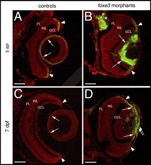
Cell proliferation in the foxe3 morphant lens. Frozen sections from control and foxe3 morpholino-injected embryos were immunolabeled for PCNA, a marker for proliferating cells. The control lens exhibits very few PCNA-positive cells at 5 and 7 dpf (panels A and C, respectively; arrows). Large numbers of anti-PCNA-labeled cells are identified in the foxe3 morphant lens at these ages (panels B and D, arrows). Small groups of PCNA-expressing cells are identified in the retinal circumferential marginal zones of both control and morphant eyes (panels A–D, arrowheads). A region of PCNA-labeled cells extends from the iris across the lens in the 7 dpf foxe3 morphant (panel D, double arrow). In addition, a region of cell proliferation in the brain is identified in the 5 dpf foxe3 morphant section (panel B, asterisk). Scale bars = 50 μm. Abbreviations: PL, photoreceptor layer; INL, inner nuclear layer and GCL, ganglion cell layer.

Expression Data
Gene:
Fish:
Knockdown Reagent:
Anatomical Term:
Stage Range: Day 5 to Days 7-13

Expression Detail
Antibody Labeling
Phenotype Data
Fish:
Knockdown Reagent:
Observed In:
Stage Range: Day 5 to Days 7-13

Phenotype Detail
Acknowledgments
This image is the copyrighted work of the attributed author or publisher, and ZFIN has permission only to display this image to its users. Additional permissions should be obtained from the applicable author or publisher of the image.

Reprinted from Mechanisms of Development, 123(10), Shi, X., Luo, Y., Howley, S., Dzialo, A., Foley, S., Hyde, D.R., and Vihtelic, T.S., Zebrafish foxe3: Roles in ocular lens morphogenesis through interaction with pitx3, 761-782, Copyright (2006) with permission from Elsevier. Full text @ Mech. Dev.