
Fig. 4
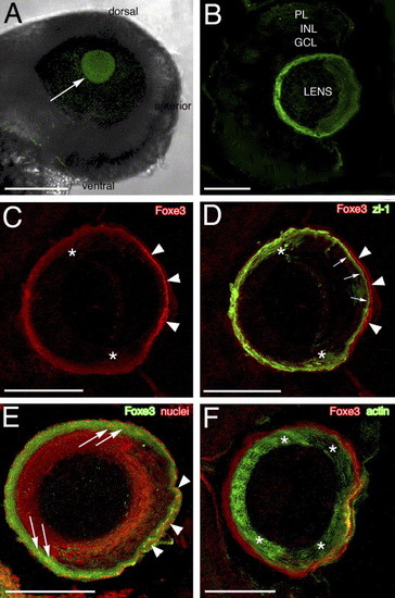
Foxe3 immunolocalization. The purified Foxe3 antiserum was used to localize protein expression in the lens by whole-mount labeling of wild-type embryos (panel A) and 4 and 5 dpf frozen larval eye sections (panels B–F). The Foxe3 protein is detected in the lens at 48 hpf (panel A, arrow). On frozen eye sections at 4 dpf, the majority of Foxe3 protein in the lens is localized to the most peripheral cells and the protein is not detected in the retina (panel B). The double immunolocalization of Foxe3 and zl-1 is shown in panels C and D. The Foxe3 signal (panel C, red) is most intense in the region of the distal (anterior) epithelial cells (arrowheads) with more diffuse labeling in the region corresponding to the cortical fibers and the elongating fiber cells (asterisks). In the overlay of the Foxe3 and zl-1 patterns (panel D), zl-1 expression (green) is detected in the elongating fiber cells (asterisks) and at the base of the distal epithelial cells (arrows). Foxe3 expression in the distal epithelial cells (arrowheads) does not overlap with the strong zl-1 signal in the cortical fibers at the base of these epithelial cells (arrows). In panel E, the anti-Foxe3 labeling (green) overlaps with propidium iodide-stained lens cell nuclei (red) in the region of the elongating fiber cells (arrows) and the epithelial cells (arrowheads). In comparison, Foxe3 expression (red) does not overlap with the majority of the actin localized in the differentiated fiber cells (green) of the lens cortex (panel F, asterisks). Scale bars = 125 μm (panel A) or 50 μm (panels B–F). Abbreviations: PL, photoreceptor layer; INL, inner nuclear layer and GCL, ganglion cell layer. |
| Gene: | |
|---|---|
| Fish: | |
| Anatomical Terms: | |
| Stage Range: | Long-pec to Day 4 |
Reprinted from Mechanisms of Development, 123(10), Shi, X., Luo, Y., Howley, S., Dzialo, A., Foley, S., Hyde, D.R., and Vihtelic, T.S., Zebrafish foxe3: Roles in ocular lens morphogenesis through interaction with pitx3, 761-782, Copyright (2006) with permission from Elsevier. Full text @ Mech. Dev.