
Fig. 8
- ID
- ZDB-IMAGE-070822-7
- Genes
- Publication
- Shi et al., 2006 - Zebrafish foxe3: Roles in ocular lens morphogenesis through interaction with pitx3
- All Figures
- Figures for Shi et al., 2006
|
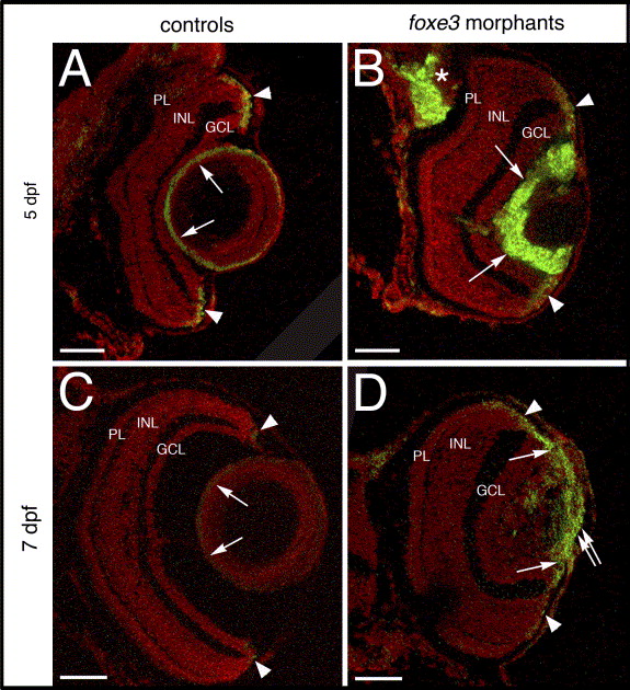
Fig. 8 Cell proliferation in the foxe3 morphant lens. Frozen sections from control and foxe3 morpholino-injected embryos were immunolabeled for PCNA, a marker for proliferating cells. The control lens exhibits very few PCNA-positive cells at 5 and 7 dpf (panels A and C, respectively; arrows). Large numbers of anti-PCNA-labeled cells are identified in the foxe3 morphant lens at these ages (panels B and D, arrows). Small groups of PCNA-expressing cells are identified in the retinal circumferential marginal zones of both control and morphant eyes (panels A–D, arrowheads). A region of PCNA-labeled cells extends from the iris across the lens in the 7 dpf foxe3 morphant (panel D, double arrow). In addition, a region of cell proliferation in the brain is identified in the 5 dpf foxe3 morphant section (panel B, asterisk). Scale bars = 50 μm. Abbreviations: PL, photoreceptor layer; INL, inner nuclear layer and GCL, ganglion cell layer.
Reprinted from Mechanisms of Development, 123(10), Shi, X., Luo, Y., Howley, S., Dzialo, A., Foley, S., Hyde, D.R., and Vihtelic, T.S., Zebrafish foxe3: Roles in ocular lens morphogenesis through interaction with pitx3, 761-782, Copyright (2006) with permission from Elsevier. Full text @ Mech. Dev.