
Fig. 7
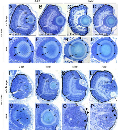
Histology of the foxe3 morphant eyes. Plastic resin-embedded sections of controls (panels A–H) and foxe3 morphants (panels I–P) were stained with methylene blue/azure II. At 3 and 5 dpf, the wild-type retina is laminated into three nuclear layers (panels A and C, uiControl). High magnification images (panels E and G) demonstrate the wild-type lens organization is characterized by a single layer of epithelial cell nuclei (arrowheads) surrounding the central lens fiber cells. Nuclei of elongating fibers are identified near the proximal lens pole (arrows). The retinal and lens morphologies of the mismatch control morpholino-injected (mmControl, panels B, D, F and H) embryos did not differ significantly from the wild-type eye morphology. Sections of the foxe3 morphant eyes at 3, 4, 5 and 7 dpf are shown in panels I–L with higher magnification views of morphant lenses shown in panels M-P. At 3 dpf (panels I and M), the morphant lens epithelial cell nuclei are enlarged, disorganized and multilayered (arrows). In addition, some fiber cells are swollen with retained nuclei (asterisks). These epithelial cell abnormalities are also evident at 4 dpf (panels J and N, arrowheads). At 5 dpf, the morphant lens exhibits a more homogenous appearing population of nucleated cells (panels K and O) with a relatively small fiber cell mass (panel K, arrow). At 7 dpf, the nucleated cells of the morphant lens appear organized into layers (panels L and P, asterisks). Some morphant lens epithelial cells also exhibit pigmentation at 5 and 7 dpf (panels O and P, arrowheads). The morphant lenses shown at high magnification in panels M, O and P are from different morphant eyes than those shown in panels I, K and L, while the lens in panel N is the same as shown at lower magnification in panel J. Scale bars = 50 μm. Abbreviations: PL, photoreceptor layer; INL, inner nuclear layer; GCL, ganglion cell layer; OpN, optic nerve; uiControl, uninjected control and mmControl, mismatch morpholino-injected control. |
| Fish: | |
|---|---|
| Knockdown Reagent: | |
| Observed In: | |
| Stage Range: | Protruding-mouth to Days 7-13 |
Reprinted from Mechanisms of Development, 123(10), Shi, X., Luo, Y., Howley, S., Dzialo, A., Foley, S., Hyde, D.R., and Vihtelic, T.S., Zebrafish foxe3: Roles in ocular lens morphogenesis through interaction with pitx3, 761-782, Copyright (2006) with permission from Elsevier. Full text @ Mech. Dev.