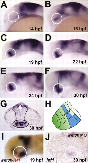
lef1 is expressed in the posterior hypothalamus during embryonic development. Lateral views are shown with anterior towards the left. White circles outline posterior hypothalamus. (A) At 14 hpf, lef1 is strongly expressed in the dorsal midbrain, but does not show specific expression in the developing hypothalamus. (B-F) At 16 hpf, lef1 expression first appears in the presumptive posterior hypothalamus, and this expression is maintained through 30 hpf. After 19 hpf, expression is present in dorsal and ventral regions of the posterior hypothalamus. Black line in F indicates plane of section in G. (G) Transverse section through the posterior hypothalamus (black oval) at 30 hpf, showing lef1 expression in both the medial mitotic cells and the lateral postmitotic cells of the posterior region. (H) Schematic depiction of lef1 expression domain in 30 hpf zebrafish hypothalamus. Numbered regions are based on those of Hauptmann and Gerster (Hauptmann and Gerster, 2000), and lef1 expression is shown in blue. (I) At 19 hpf, wnt8b (blue) and lef1 (red) show overlapping and adjacent expression in the posterior hypothalamus. (J) In wnt8b morphants, lef1 expression is reduced throughout the brain.
|
