
Fig. 6
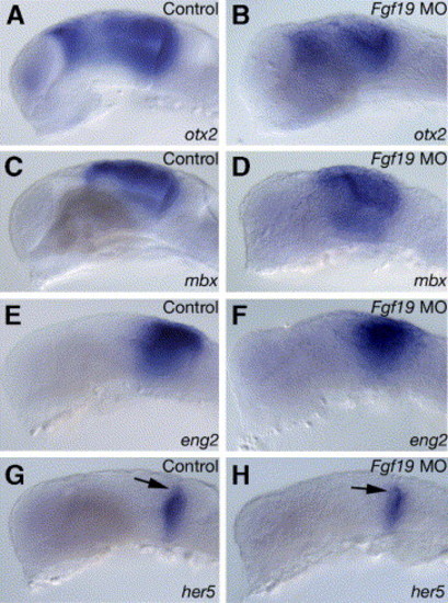
Expression of region-specific markers in the midbrain and cerebellum in the Fgf19 MO-injected embryos at 25 hpf. Embryos were injected with control Fgf19 MO (A, C, E and G) and Fgf19 MO (B, D, F and H). (A and B) In the Fgf19 MO-injected embryos, otx2 expression was detected in the dorsal thalamus and midbrain, while the expression domains were smaller in the Fgf19 MO-injected embryos than in the control Fgf19 MO-injected embryos. Furthermore, otx2 expression in the olfactory epithelium was lost. (C–F) mbx (C and D) and eng2 (E and F) were expressed in similar regions in the control Fgf19 MO-injected and Fgf19 MO-injected embryos, while the expression domains were smaller in the Fgf19 MO-injected embryos than in the control Fgf19 MO-injected embryos. (G and H) Expression of her5 in the MHB was normal in the Fgf19 MO-injected embryos. Lateral views with anterior to the left and dorsal to the top. Arrows in panels G and H indicate the MHB. |
| Genes: | |
|---|---|
| Fish: | |
| Knockdown Reagent: | |
| Anatomical Terms: | |
| Stage: | Prim-5 |
Reprinted from Developmental Biology, 288(1), Miyake, A., Nakayama, Y., Konishi, M., and Itoh, N., Fgf19 regulated by Hh signaling is required for zebrafish forebrain development, 259-275, Copyright (2005) with permission from Elsevier. Full text @ Dev. Biol.