
Fig. 4
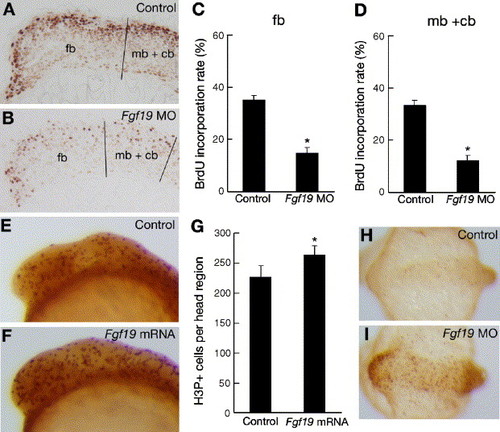
Comparison of cell proliferation and cell death patterns in control embryos and Fgf19 MO- or Fgf19 mRNA-injected embryos. (A and B) Embryos injected with control Fgf19 MO (A) and Fgf19 MO (B) were labeled with BrdU and stained using an anti-BrdU antibody. Lateral views with anterior to the left and dorsal to the top. (A) BrdU-positive cells (light brown in A) were mostly detected in the dorsal region of the brain in the control embryos at 16 hpf. (B) Representative sagittal sections of the head region of stained embryos that show fewer labeled cells (light brown in B) in the Fgf19 MO-injected embryos. (C and D) The proportions of BrdU-positive cells in the forebrain (C) and the midbrain and cerebellum (D) of the control Fgf19 MO-injected and Fgf19 MO-injected embryos were quantitatively examined by counting BrdU-positive and -negative cells in the sections. Results are the means ± SD for three independent sections from three embryos. Asterisks indicate statistical significance compared with the control (*P < 0.005). The forebrain (fb), and the midbrain and cerebellum (mb + cb) regions, which we defined in the sections, are separated by black lines. (E and F) Wild-type embryos (E) and embryos injected with Fgf19 mRNA (F) were stained using an anti-H3P antibody. Lateral views with anterior to the left and dorsal to the top. Increased numbers of H3P-positive cells (dark brown in panel F) were detected in the head region of the Fgf19 mRNA-injected embryos compared with the wild-type embryos at 16 hpf. (G) Numbers of H3P-positive cells in the forebrain, midbrain and cerebellum of the wild-type and Fgf19 mRNA-injected embryos were counted. Results are the means ± SD for four embryos. Asterisks indicate statistical significance compared with the wild type (*P < 0.05). (H and I) Apoptotic cells in the wild-type embryos (H) and embryos injected with Fgf19 MO (I) were marked by TUNEL staining. Dorsal views with anterior to the left. Substantially, increased numbers of apoptotic cells (light brown in I) were detected in the head region of the Fgf19 MO-injected embryos at 16 hpf. |
Reprinted from Developmental Biology, 288(1), Miyake, A., Nakayama, Y., Konishi, M., and Itoh, N., Fgf19 regulated by Hh signaling is required for zebrafish forebrain development, 259-275, Copyright (2005) with permission from Elsevier. Full text @ Dev. Biol.