FIGURE
            Fig. 6
                
                    
                        Fig. 6


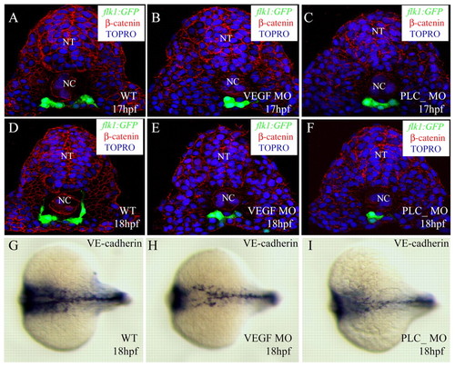
| Cell number is not crucial for angioblast migration. Transverse sections of embryos with compromised VEGF signaling visualized for GFP (green), ß-catenin (red) and TOPRO (blue) (A-F), and in situ hybridization with VE-cadherin (cdh5) (G-I). The sections shown are at the level of the 6th somite (A-F). Control embryos (A,D,G), vegf MO-injected embryos (B,E,H), plcg1 MO-injected embryos (C,F,I). The angioblasts in embryos with compromised VEGF signaling are in a similar position to those in control embryos, while the numbers of angioblasts are significantly reduced. NT, neural tube; NC, notochord. | 
                
                    
                        Expression Data


    | Gene: | |
|---|---|
| Fish: | |
| Knockdown Reagents: | |
| Anatomical Term: | |
| Stage: | 14-19 somites | 
                
                    
                        Expression Detail


                        Antibody Labeling


                        Phenotype Data


                        Phenotype Detail


                        Acknowledgments


This image is the copyrighted work of the attributed author or publisher, and
ZFIN has permission only to display this image to its users.
Additional permissions should be obtained from the applicable author or publisher of the image.
      Full text @ Development


        