
Fig. 8
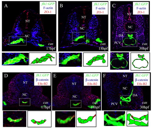
Junction formation and arterial endothelial cell differentiation in endodermless embryos. Transverse sections of endodermless embryos. Tg(flk1:EGFP)s843;cas mutants visualized for: (A-C) GFP (green), filamentous actin (blue) and ZO1 (red); and (D-F) GFP (green) ephrin B2 (red) and ß-catenin (blue). The sections shown are at the level of the 6th (A,D), 10th (B,E), and 14th somites (C,F). The outlined areas are magnified and shown with schematic drawings. Despite the absence of endoderm, differentiation of angioblasts into arterial and venous endothelial cells occurs as in wild-type embryos. NT, neural tube; NC, notochord; DA, dorsal aorta; PCV, posterior cardinal vein. |