
Fig. 6
- ID
- ZDB-IMAGE-051214-6
- Genes
- Publication
- Jin et al., 2005 - Cellular and molecular analyses of vascular tube and lumen formation in zebrafish
- All Figures
- Figures for Jin et al., 2005
|
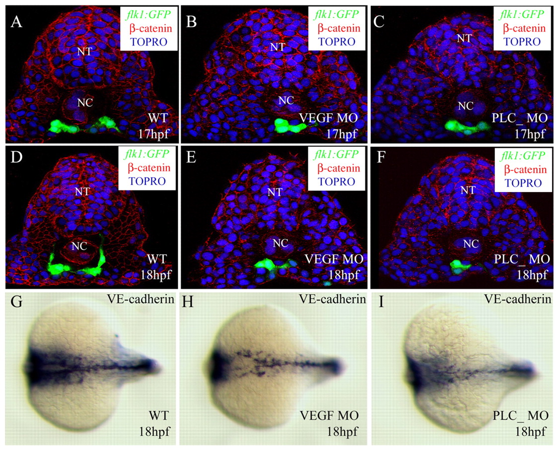
Fig. 6 Cell number is not crucial for angioblast migration. Transverse sections of embryos with compromised VEGF signaling visualized for GFP (green), ß-catenin (red) and TOPRO (blue) (A-F), and in situ hybridization with VE-cadherin (cdh5) (G-I). The sections shown are at the level of the 6th somite (A-F). Control embryos (A,D,G), vegf MO-injected embryos (B,E,H), plcg1 MO-injected embryos (C,F,I). The angioblasts in embryos with compromised VEGF signaling are in a similar position to those in control embryos, while the numbers of angioblasts are significantly reduced. NT, neural tube; NC, notochord.