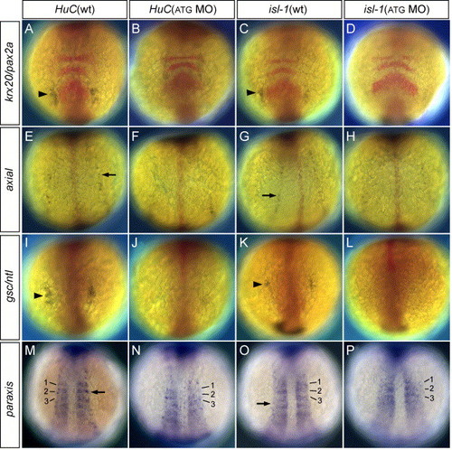
Double in situ hybridization of HuC/isl-1 and neural/mesodermal markers in MOs-injected embryos. (A, C, E, G, I, K, M, O) Wild-type embryos at 3-somite stage. (B, D, F, H, J, L, N, P) 20 ng ATG-dlx3b-MO/20 ng ATG-dlx4b-MO-injected embryos at 3-somite stage. (A and B) Expression of HuC (blue) and krx20/pax2a, a marker of rhombomeres 3 and 5 and midbrain–hindbrain boundary (red). (C and D) Expression of isl-1 (blue) and krx20/pax2a (red). (E and F) Expression of HuC (blue) and axial marking floor plate (red). (G and H) Expression of isl-1 (blue) and axial (red). (I and J) Expression of HuC (blue) and gsc/ntl expressed in the prechordal plate and notochord (red). (K and L) Expression of isl-1 (blue) and gsc/ntl (red). (M and N) Expression of HuC (blue) and paraxis showing expression in the somites (blue). Numbers 1–3 correspond to the somites. (O and P) Expression of isl-1 (blue) and paraxis (blue). Numbers 1–3 correspond to the somites. In (B, D, J, L), neurons in the trigeminal placodes (arrowhead) are highly reduced or absent, shown by the expression of HuC and isl-1. The expression of HuC and isl-1 in the RB neurons (F, H, N, P; arrow) are highly reduced or absent. However, no changes are observed in neural/mesodermal marker expression in dlx3b and dlx4b MOs-injected embryos (B, D, F, H, J, L, N, P).
|
