|
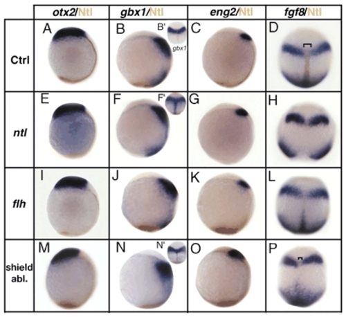
Expression of MHB markers is normal in mutants with defects in notochord formation and in shield-ablated embryos. (A-D) In situ hybridization and Ntl immunostaining (brown) of control embryos, (E-H) ntl mutant embryos, (I-L) flh mutant embryos and (M-P) shield-ablated embryos at tailbud stage. Probes were applied: otx2 (A,E,I,M), gbx1 (B,F,J,N), eng2 (C,G,K,O), fgf8 (D,H,L,P). In B', no Ntl immunostaining has been performed. No changes in the AP position of the different MHB markers analyzed is observed in ntl mutants, in flh mutants or in shield-ablated embryos when compared with wild-type embryos. (D,P) Brackets indicate the reduced midline width following shield ablation. (A-O) Lateral views, anterior upwards; (D,H,L,P,B',F',N') dorsal views, anterior upwards.
|