
FIGURE
Fig. 5
Fig. 5
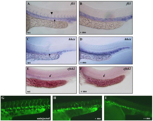
Angiogenesis is disrupted in scl morphants. Transcripts for fli1 (A, B), hhex (C, D), and efnb2 (E, F) in uninjected and scl morphant embryos. In A, the arrowhead indicates the expression in intersomitic vessels, and in A, C, E–F, the arrow indicates expression in the dorsal aorta. G) Uninjected Tg(lmo2:EGFP) embryos, showing fluorescence in the axial and intersomitic vessels and blood cells. In the presence of increasing amounts scl morpholino (H, I), increased disruption of angiogenesis occurred. All images are lateral views of trunk region, anterior to the left. |
Expression Data
| Genes: | |
|---|---|
| Fish: | |
| Knockdown Reagents: | |
| Anatomical Terms: | |
| Stage: | Prim-5 |
Expression Detail
Antibody Labeling
Phenotype Data
Phenotype Detail
Acknowledgments
This image is the copyrighted work of the attributed author or publisher, and
ZFIN has permission only to display this image to its users.
Additional permissions should be obtained from the applicable author or publisher of the image.
Reprinted from Developmental Biology, 277(2), Dooley, K.A., Davidson, A.J., and Zon, L.I., Zebrafish scl functions independently in hematopoietic and endothelial development, 522-536, Copyright (2005) with permission from Elsevier. Full text @ Dev. Biol.