
- Title
-
Assessment of Oral Albendazole and Fumagillin in the Treatment of Pseudoloma neurophilia in Adult Zebrafish
- Authors
- Lavin, E.S., Getchell, R.G., Daugherity, E.K., Kent, M.L., Frosolone, A.D., Ivanek, R.
- Source
- Full text @ Comp. Med.
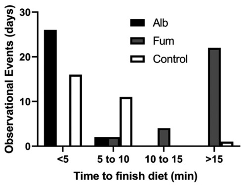
For the 28-d pilot study, each study group was observed daily and the time required to finish the diet recorded. On most days, the albendazole group took less than 5 min to finish the diet (no feed observed in the tank after 5-min post-feeding), whereas on most days the fumagillin group took over 15 min to finish the diet. All groups finished the diet by 60-min post-feeding. Fisher exact test was used for statistical analysis where albendazole and fumagillin were each compared to the control and post hoc analysis using a Benjamini-Hochberg correction. A P value < 0.05 was considered significant. |
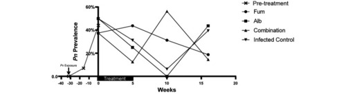
The left side of the Y axis shows Pn prevalence following Pn exposure. At the beginning of the experiment (week 0), overall Pn prevalence across groups was 44%. The right side of the Y axis demonstrates Pn prevalence for each experimental group over time following treatment. The Pn prevalence obtained for the uninfected control group was 0 at all time points, and this group is omitted from this figure. |
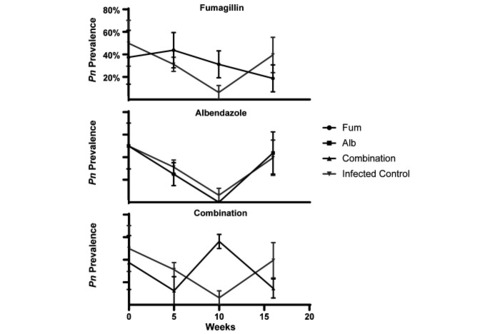
Plots demonstrating Pn prevalence over time as determined by whole-body qPCR at 5, 10, and 16-wk time points. The figure shows predicted means and associated standard error bars from raw data. Prevalence of the infected control group was compared to each of the treatment groups. There was no significant association between treatment group and Pn prevalence. The Pn prevalence obtained for the uninfected control group was 0 at all time points, and this group is omitted from this figure. |
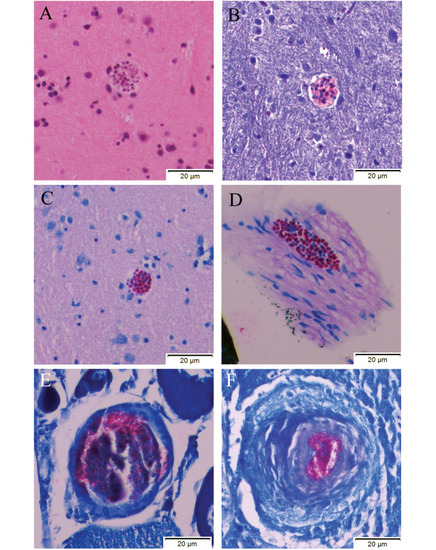
Histology results for a zebrafish that was exposed to the Pn inoculum and was found at the bottom of a tank with multifocal superficial erosions over the coelom approximately 12-mo post-exposure. This fish was not enrolled in either the pilot study or the experiment, although it had been exposed to the same inoculum as infected fish enrolled in these studies. This animal was euthanized and processed for histology. All images were obtained at 40× magnification. (A-C) Images show a Pn xenoma within the hindbrain of the fish (A) H and E, (B) Luna, (C) Fite Faraco. Image D shows another xenoma at the level of the meninges (D) Fite Faraco. Incidental granulomas containing acid-fast bacteria (suspected Mycobacterium sp.) were seen in the ovaries (E) Fite Faraco and liver (F) Fite Faraco. Zebrafish exposed to Pn in this study were reared in a facility with endemic M. chelonae identified on previous surveillance monitoring reports. |
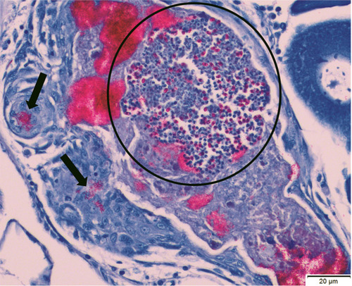
Histology results for a zebrafish that was exposed to the Pn inoculum but was not enrolled in either the pilot study or the experiment, although it had been exposed to the same inoculum as infected fish enrolled in these studies. This animal did not demonstrate any clinical signs and was euthanized 13-mo post-exposure. This histology section, stained with Fite Faraco, demonstrates numerous Pn spores within a parasite cluster (circled) in the ovary, as well as aggregates of acid-fast bacilli (arrows: suspected Mycobacterium sp). |