
|
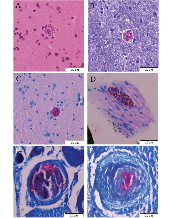
Fig. 4 Histology results for a zebrafish that was exposed to the Pn inoculum and was found at the bottom of a tank with multifocal superficial erosions over the coelom approximately 12-mo post-exposure. This fish was not enrolled in either the pilot study or the experiment, although it had been exposed to the same inoculum as infected fish enrolled in these studies. This animal was euthanized and processed for histology. All images were obtained at 40× magnification. (A-C) Images show a Pn xenoma within the hindbrain of the fish (A) H and E, (B) Luna, (C) Fite Faraco. Image D shows another xenoma at the level of the meninges (D) Fite Faraco. Incidental granulomas containing acid-fast bacteria (suspected Mycobacterium sp.) were seen in the ovaries (E) Fite Faraco and liver (F) Fite Faraco. Zebrafish exposed to Pn in this study were reared in a facility with endemic M. chelonae identified on previous surveillance monitoring reports.