
Image
|
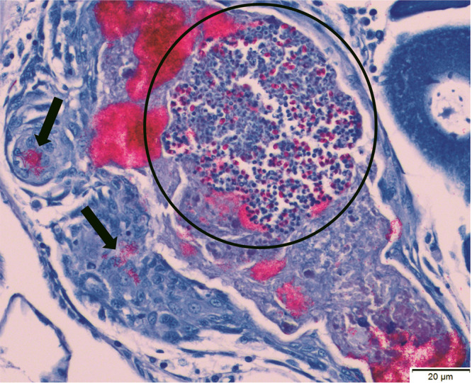
Figure Caption
Fig. 5 Histology results for a zebrafish that was exposed to the Pn inoculum but was not enrolled in either the pilot study or the experiment, although it had been exposed to the same inoculum as infected fish enrolled in these studies. This animal did not demonstrate any clinical signs and was euthanized 13-mo post-exposure. This histology section, stained with Fite Faraco, demonstrates numerous Pn spores within a parasite cluster (circled) in the ovary, as well as aggregates of acid-fast bacilli (arrows: suspected Mycobacterium sp).
Acknowledgments
This image is the copyrighted work of the attributed author or publisher, and
ZFIN has permission only to display this image to its users.
Additional permissions should be obtained from the applicable author or publisher of the image.
Full text @ Comp. Med.