
- Title
-
Wnt signaling is required for early development of zebrafish swimbladder
- Authors
- Yin, A., Korzh, S., Winata, C.L., Korzh, V., and Gong, Z.
- Source
- Full text @ PLoS One

Expression of new maker genes in different tissue layers of the zebrafish swimbladder as assayed by WISH. (A–F) Expression of sox2 in the epithelium of swimbladder at 24 hpf (A), 36 hpf (B), 48 hpf (C) and 72 hpf (D–F). Panels (A–D) are lateral view while (E,F) are cross sections of the embryo shown in (D) with the section planes indicated. (G–I) Expression of has2 in the mesenchyme layer of swimbladder at 48 hpf (G, lateral view) and 72 hpf (H, ventral view; I, cross section). (H2) Expression of fgf10a in swimbladder (ventral view) for comparison of has2 expression in (H). (J–L) Expression of hprt1l in the outer mesothelium of swimbladder at 48 hpf (J, lateral), and 72 hpf (K, ventral; L, cross section). (M–R) Expression of elovl1a in the outer mesothelium of swimbladder at 48 hpf (M, lateral; N, ventral; O, cross section) and 72 hpf (P, lateral; Q, ventral; R, cross section). Dotted red circles indicated swimbladder and yellow circles indicated epithelium. All embryos were laterally oriented with anterior to the left unless specified. Abbreviations: asb, anterior swimbladder bud; e, epithelium; g, gut; m, mesenchyme; n, notochord; o, outer mesothelium; pd, pneumatic duct; sb, swimbladder. Scale bar = 200 μm. Panel (A) scale bar applies to all whole mount images and Panel (F) scale bar is for all cross section images. |

Expression of Wnt pathway genes in zebrafish swimbladder as detected by WISH. (A–C) Expression of wnt5b in the mesenchyme of swimbladder at 36 hpf (A) and 72 hpf (B,C). (D–F) Expression of fz2 in the mesenchyme and outer mesothelium of swimbladder at 36 hpf (D) and 72 hpf (E,F). (G–I) Expression of fz7b in mesenchyme and outer mesothelium of swimbladder at 36 hpf (G) and 72 hpf (H,I). (J–L) Expression of lef1 in the mesenchyme and outer mesothelium of swimbladder at 36 hpf (J) and 72 hpf (K,L). (M–O) Expression of tcf3 in the epithelium and outer mesothelium of swimbladder at 36 hpf (M) to 72 hpf (N,O). Panels (C, F, I, L, O) are cross sections. All embryos were laterally oriented with anterior to the left unless specified. Dotted red circles indicated swimbladder and yellow circles indicated epithelium. Abbreviations: e, epithelium; g, gut; m, mesenchyme; n, notochord; o, outer mesothelium; pf, pectoral fin; pd, pneumatic duct; sb, swimbladder. Scale bars = 200 μm. Panel (A) scale bar applies to all whole mount images and Panel (C) scale bar is for all cross section images. |

Induction of GFP-fusion proteins and inhibition of Wnt signaling in the hs:Dkk1-GFP and hs:ΔTcf-GFP transgenic embryos by heat-shock treatment. (A–H) Induction of GFP-fusion proteins in hs:Dkk1-GFP and hs:ΔTcf-GFP transgenic embryos. Heat-shock was performed at various stages and live images were taken at 72 hpf. (A) Lack of GFP expression in wild type sibling after heatshock treatment. (B–D) Live image of GFP expression in hs:Dkk1-GFP embryos heat-shocked at 12 hpf (B), 24 hpf (C) and 48 hpf (D). (E–H) Live image of GFP expression in hs:ΔTcf-GFP embryos heat-shocked at 12 hpf (E), 24 hpf (F), 36 hpf (G) and 48 hpf (H). Note that the expression of GFP in hs:ΔTcf-GFP embryos (E–H) were stronger than that of hs:Dkk1-GFP embryos (A–D), implying a stronger inhibition of Wnt signaling in hs:ΔTcf-GFP embryos. (I–L) Analysis of GFP-fusion protein expression in the swimbladder of hs:ΔTcf-GFP transgenic embryos. Transgenic embryos were heat-shocked at 66 hpf and live images were taken at 72 hpf (I), followed by immunohistochemical staining using anti-GFP antibody (K,L) and DAPI counterstaining (J,L). Panels (J–L) are cross sections. (M,N) Real time RT-PCR assays of selected target genes of the Wnt signaling after heat-shock blocking Wnt signaling in hs:Dkk1-GFP (M) and hs:ΔTcf-GFP (N) transgenic embryos. Stronger inhibition of wnt signaling targets genes axin2, c-myc, cyclinD1 and lef1 in hs:ΔTcf-GFP fishes (M) than hs:Dkk1-GFP fishes (N) were observed (p<0.05). All embryos were lateral oriented with anterior to the left unless specified. Dotted white circles indicated swimbladder. Abbreviations: g, gut. Scale bars = 200 μm. |
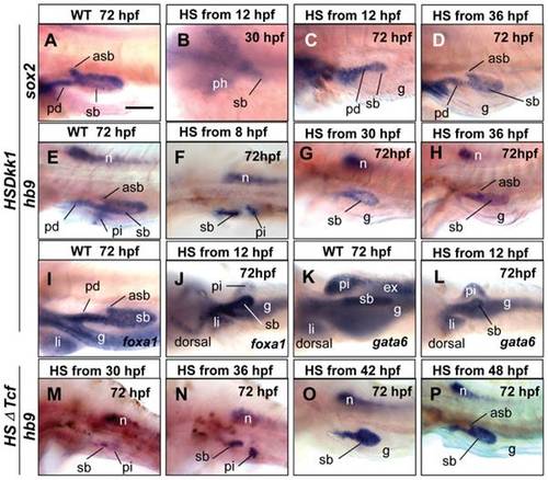
Effects of temporal inhibition of wnt signaling on the epithelium development of swimbladder. (A–L) Expression of marker genes in hs:Dkk1-GFP embryos after heat-shock treatment at various stages. (A–D) Expression of sox2 in swimbladder epithelium in wild type sibling at 72 hpf (A) and in hs:Dkk1-GFP embryos heat-shocked from different developmental stages as indicated (B–D). Expression of sox2 in the swimbladder epithelium anlage at 30 hpf when heat-shocked from 12 hpf (B). (E–H) Expression of hb9 at 72 hpf in wild type swimbladder epithelium, in 72 hpf hs:Dkk1-GFP larvae heat-shocked from 8 hpf, 30 hpf (G) and 36 hpf (H). (I, J) Expression of foxA1 at 72 hpf in the epithelium of endoderm organs in wild type and transgenic larvae heat-shocked from 12 hpf (J). (K, L) Expression of gata6 in the epithelium of endoderm organs at 72 hpf in wild type and transgenic fishes heat-shocked from 12 hpf. (M–P) Expression of hb9 in the epithelium of swimbladder at 72 hpf in transgenic larvae heat-shocked from 30 hpf, 36 hpf, 42 hpf and 48 hpf. Note the presence of anterior swimbladder bud (asb) in (D, H, P). All embryos were laterally oriented with anterior to the left unless specified. Abbreviations: asb, anterior swimbladder bud; ex, exocrine pancreas; g, gut; li, liver; n, notochord; pd, pneumatic duct; ph, pharynx; pi, pancreatic islet; sb, swimbladder. Scale bar in (A) = 200 μm for all panels. |

Effects of temporal inhibition of Wnt signaling on swimbladder mesenchyme and smooth muscles. (A, C) Expression of has2 in swimbladder mesenchyme at 72 hpf in wild type and hs:Dkk1-GFP fishes that were heat-shocked from 12 hpf. (B) Absence of has2 expression at 48 hpf in the putative swimbladder when hs:Dkk1-GFP larvae were heat-shocked from 12 hpf. (D) Expression of has2 in the size-reduced mesenchyme in hs:ΔTcf-GFP larvae heat-shocked from 36 hpf. (E–G) Expression of fgf10a at 72 hpf in mesenchyme of wild type (E), transgenic hs:Dkk1-GFP larvae heat-shocked from 12 hpf (F) and 60 hpf (G). (H) Expression of fgf10a in the size-reduced mesenchyme in hs:ΔTcf-GFP larvae heat-shocked from 36 hpf. (I–K) Expression of acta2 at 72 hpf in mesenchyme of wild type, hs:Dkk1-GFP larvae heat-shocked from 30 hpf and 36 hpf. Note absence of acta2 staining in (J) and the reduced size of swimbladder in (K). (L) Expression of acta2 in the size-reduced mesenchyme in hs:ΔTcf-GFP larvae heat-shocked from 48 hpf. Scale bar in (A) = 200 μm for all panels. |
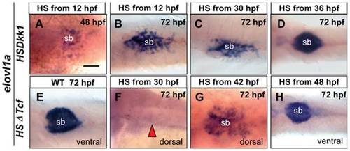
Effects of temperal inhibition of Wnt signaling on swimbladder mesothelium development. (A, B) Expression of elovl1a in outer mesothelium at 48 hpf and 72 hpf when hs:Dkk1-GFP fishes were heat-shocked from 12 hpf. The elovl1a expressing cells were disorganized at 72 hpf when larvae were heat-shocked from 24 hpf (C), but were well organized with smaller size when larvae were heat-shocked from 30 hpf (D). (E) Expression of elovl1a in wild-type outer mesothelium. The elovl1a expressing cells in the outer mesothelium at 72 hpf were absent when hs:ΔTcf-GFP larvae were heat-shocked from 30 hpf (F), were present but disorganized when larvae were heat-shocked from 42 hpf (G), and were properly organized but with smaller size when fishes were heat-shocked from 42 hpf (H). All embryos were laterally oriented with anterior to the left unless specified. Red arrows indicated absence of swimbladder at putative locations. Abbreviations: g, gut; sb, swimbladder. Scale bar in (A) = 200 μm for all panels. |

Effects of Wnt inhibition on cell proliferation in swimbladder. hs:ΔTcf-GFP fish was out-crossed with AB wild type fish, the resultant heterozygous embryos and their wild type siblings were heat-shocked at 66 hpf and fixed at 72 hpf for proliferation assay. (A–F) Proliferation assay detecting PCNA-positive cells (red) with DAPI counterstaining (green). The number of PCNA-positive cells (red) was greatly reduced in transgenic fishes (D–F) (n = 5) compared to that of controls (A–C) (n = 5). (G–L) Staining for phosphorylated histone H3 (PH3, red) with DAPI counterstaining (green). Compared to wild type fishes (G–I) (n = 5), the number of PH3-positive cells (red) was greatly reduced in transgenic fishes (D–F) (n = 5). Dotted white circles indicated swimbladder. Abbreviation: g, gut. Scale bar = 200 μm for all panels. |
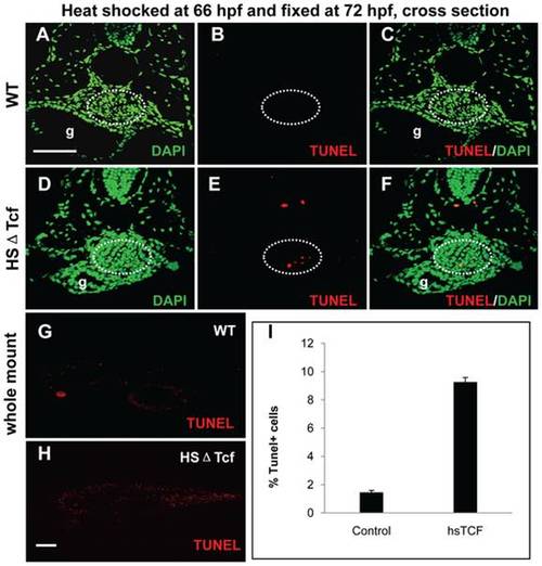
Effects of Wnt inhibition on cell apoptosis in swimbladder. hs:ΔTcf-GFP heterozygous larvae and their wild type siblings were heat-shocked at 66 hpf and fixed at 72 hpf for TUNEL assay. (A–F) TUNEL-positive cells (red) and DAPI-stained cells (green) shown in cross section. The number of TUNEL-positive cells in swimbladder greatly increased in transgenic fishes (D–E) (n = 11 of 13, 85%) compared to that of controls (A–C) (n = 9 of 10, 90%). (G, H) Imaging of whole mount TUNEL-stained (red) transgenic fishes (H) and controls (G). (I) The statistics assays showed that the percentage of apoptosis cells in heat-shocked transgenics increased from 1.45% to 9.26% (I) (p = 0.01). Note the globally increased number of TUNEL-positive cells in (H). All embryos were laterally oriented with anterior to the left. Dotted white circles indicated swimbladder. Abbreviation: g, gut. Scale bar = 200 μm for all panels. |

Crosstalk of Wnt and Hh signalling in swimbladder development. (A–C) Expression of shh (A), ihh (B) and ptc1 (C) in swimbladder of controls at 72 hpf. These controls were heat-shocked at 66 hpf and fixed at 72 hpf prior to WISH. (D–F) Decreased expression of shh (D), ihh (E) and ptc1 (F) in swimbladder of hs:Dkk1-GFP fry after heat-shock. (G–I) Expression of axin1 in wild type (G), smob641 (H) and syut4 (I) at 72 hpf. (J–L) Expression of axin2 in wild type (J), smob641 (K) and syut4 (L) at 72 hpf. Embryos in (G–L) were short-stained for the same one hour for comparison and no detectable axin1 and axin2 signals were observed in controls but prolonged staining (5 hours) showed enriched hybridization signals in the swimbladder in controls (G′ and J′). Compared to the short-staining pictures in (G) and (J), increased global expression of axin1 and axin2 in smob641 and syut4 was observed although the swimbladder development has been severely affected in the two mutants [26]. Red arrows indicate the position of swimbladder. Abbreviation: sb, swimbladder. Scale bar in (A) = 200 μm for all panels. |
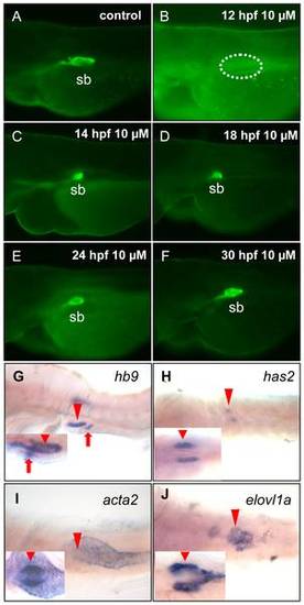
Chemical inhibition of Wnt signaling and its effect on swimbladder development. (A–F) Effect of Wnt inhibition on development of swimbladder epithelia. Et(krt4:EGFP)sq33-2 embryos were doped with 10 µM IWR-1 from various time points and then live-imaged at 72 hpf. Panel (A) shows normal swimbladder epithelium in a controls without IWE-1 treatment and panels (B–F) show the lack of swimbladder epithelium in embryos treated from 12 hpf and smaller swimbladders in embryos treated from 14 hfp (C), 16 hpf (D), 24 hpf (E) and 30 hpf (F). Dotted circle indicates the position of swimbladder. (G–J) Effect of Wnt signaling inhibition on development of different tissue layers of swimbladder. Control embryos were treated with 10 µM IWR-1 from 14 hpf and were assayed by WISH at 72 hpf. The swimbladder epithelium, mesenchyme, smooth muscle and mesothelium were marked by hb9 (G) , has2 (H), acta2 (I) and elvol1a (J) expression respectively. Inserted boxes present their expression in normally developed swimbladder in controls for comparison. (G) shows lateral view and (H–J) show ventral view. The red arrowheads indicate the swimbladder, whereas the red arrows indicate pancreas. Abbreviation: sb, swimbladder. |
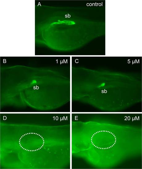
Dosage-dependent effect of IWR-1 on specification of the swimbladder epithelial cells. The Et(krt4:EGFP)sq33-2 embryos were cultured in egg water with IWR-1 addition from 12 hpf at a concentration as indicated, and was live-imaged for GFP fluorescence at 72 hpf. (A) Fully developed epithelium of the swimbladder at 72 hpf in a control embryo. (B, C) The small bud of the swimbladder epithelium in embryos treated with 1 μM (B) and 5 µM (C) IWR-1. (D, E) The absence of the swimbladder epithelium at 72 hpf in embryos treated with 10 μM (D) and 20 μM (E) IWR-1. Dotted circles indicate the position of swimbladder. Abbreviations: sb, swimbladder. |