
|
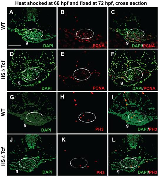
Fig. 7 Effects of Wnt inhibition on cell proliferation in swimbladder.
hs:ΔTcf-GFP fish was out-crossed with AB wild type fish, the resultant heterozygous embryos and their wild type siblings were heat-shocked at 66 hpf and fixed at 72 hpf for proliferation assay. (A–F) Proliferation assay detecting PCNA-positive cells (red) with DAPI counterstaining (green). The number of PCNA-positive cells (red) was greatly reduced in transgenic fishes (D–F) (n = 5) compared to that of controls (A–C) (n = 5). (G–L) Staining for phosphorylated histone H3 (PH3, red) with DAPI counterstaining (green). Compared to wild type fishes (G–I) (n = 5), the number of PH3-positive cells (red) was greatly reduced in transgenic fishes (D–F) (n = 5). Dotted white circles indicated swimbladder. Abbreviation: g, gut. Scale bar = 200 μm for all panels.