
|
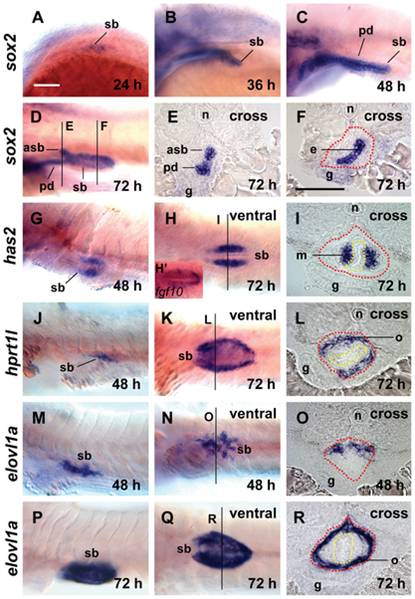
Fig. 1 Expression of new maker genes in different tissue layers of the zebrafish swimbladder as assayed by WISH.
(A–F) Expression of sox2 in the epithelium of swimbladder at 24 hpf (A), 36 hpf (B), 48 hpf (C) and 72 hpf (D–F). Panels (A–D) are lateral view while (E,F) are cross sections of the embryo shown in (D) with the section planes indicated. (G–I) Expression of has2 in the mesenchyme layer of swimbladder at 48 hpf (G, lateral view) and 72 hpf (H, ventral view; I, cross section). (H2) Expression of fgf10a in swimbladder (ventral view) for comparison of has2 expression in (H). (J–L) Expression of hprt1l in the outer mesothelium of swimbladder at 48 hpf (J, lateral), and 72 hpf (K, ventral; L, cross section). (M–R) Expression of elovl1a in the outer mesothelium of swimbladder at 48 hpf (M, lateral; N, ventral; O, cross section) and 72 hpf (P, lateral; Q, ventral; R, cross section). Dotted red circles indicated swimbladder and yellow circles indicated epithelium. All embryos were laterally oriented with anterior to the left unless specified. Abbreviations: asb, anterior swimbladder bud; e, epithelium; g, gut; m, mesenchyme; n, notochord; o, outer mesothelium; pd, pneumatic duct; sb, swimbladder. Scale bar = 200 μm. Panel (A) scale bar applies to all whole mount images and Panel (F) scale bar is for all cross section images.