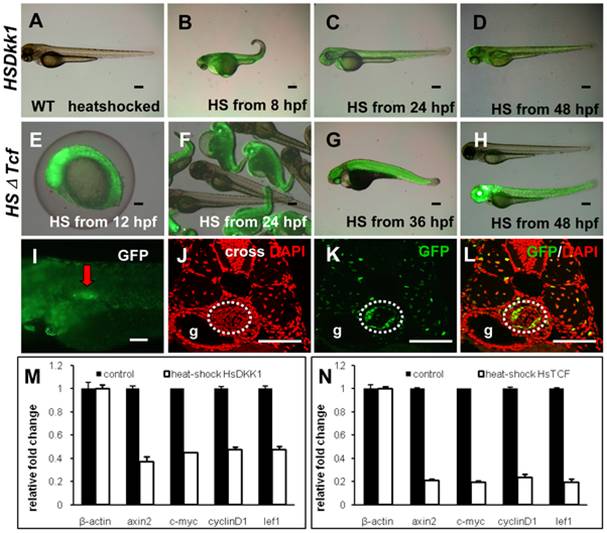
Fig. 3 Induction of GFP-fusion proteins and inhibition of Wnt signaling in the hs:Dkk1-GFP and hs:ΔTcf-GFP transgenic embryos by heat-shock treatment.
(A–H) Induction of GFP-fusion proteins in hs:Dkk1-GFP and hs:ΔTcf-GFP transgenic embryos. Heat-shock was performed at various stages and live images were taken at 72 hpf. (A) Lack of GFP expression in wild type sibling after heatshock treatment. (B–D) Live image of GFP expression in hs:Dkk1-GFP embryos heat-shocked at 12 hpf (B), 24 hpf (C) and 48 hpf (D). (E–H) Live image of GFP expression in hs:ΔTcf-GFP embryos heat-shocked at 12 hpf (E), 24 hpf (F), 36 hpf (G) and 48 hpf (H). Note that the expression of GFP in hs:ΔTcf-GFP embryos (E–H) were stronger than that of hs:Dkk1-GFP embryos (A–D), implying a stronger inhibition of Wnt signaling in hs:ΔTcf-GFP embryos. (I–L) Analysis of GFP-fusion protein expression in the swimbladder of hs:ΔTcf-GFP transgenic embryos. Transgenic embryos were heat-shocked at 66 hpf and live images were taken at 72 hpf (I), followed by immunohistochemical staining using anti-GFP antibody (K,L) and DAPI counterstaining (J,L). Panels (J–L) are cross sections. (M,N) Real time RT-PCR assays of selected target genes of the Wnt signaling after heat-shock blocking Wnt signaling in hs:Dkk1-GFP (M) and hs:ΔTcf-GFP (N) transgenic embryos. Stronger inhibition of wnt signaling targets genes axin2, c-myc, cyclinD1 and lef1 in hs:ΔTcf-GFP fishes (M) than hs:Dkk1-GFP fishes (N) were observed (p<0.05). All embryos were lateral oriented with anterior to the left unless specified. Dotted white circles indicated swimbladder. Abbreviations: g, gut. Scale bars = 200 μm.

