
- Title
-
Formation of the asymmetric pineal complex in zebrafish requires two independently acting transcription factors
- Authors
- Snelson, C.D., Burkart, J.T., and Gamse, J.T.
- Source
- Full text @ Dev. Dyn.
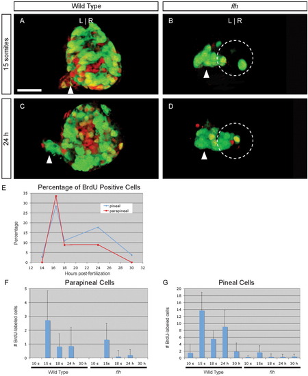
Cell division of parapineal precursors is largely complete by 18 s. A-D: Dorsal views of 4 d larvae, with BrdU (red) and foxd3:gfp (green) labeling in the epithalamus, following a BrdU pulse at (A, B) 15 s or (C, D) 24 hr stage in (A, C) WT or (B, D) flh mutant embryos. Dashed circles in B, D indicate the pineal cells. Scale bar = 25 μM. E: Line graph of the percentage of WT parapineal (red line) or pineal (blue line) cells that are labeled at 4 d, following a BrdU pulse at the indicated developmental stages. F: Bar graph showing the absolute number of BrdU-positive parapineal cells in WT or flh embryos. G: BrdU-positive pineal cells in WT or flh embryos. EXPRESSION / LABELING:
|
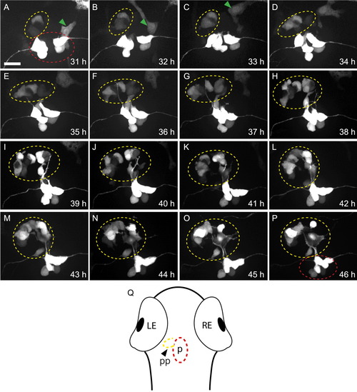
Parapineal cells migrate asymmetrically in flh mutants. All panels are dorsal views of foxd3:gfp labeling in the epithalamus of a single embryo at the times indicated. Parapineal cells are circled in yellow, pineal cells in red; a neural crest cell (which also expresses foxd3:gfp) in A-C is indicated by a green arrowhead. A: The first labeled parapineal cells are apparent near the midline at 31 hr. B-E: They migrate leftward. F-P: They are joined by more leftward-migrating parapineal cells during the subsequent 10 hr. Scale bar = 25 μm. Q: Diagram of a zebrafish larvae, showing the relative position of the pineal (p, red oval) and parapineal (pp, yellow oval) within the head. |
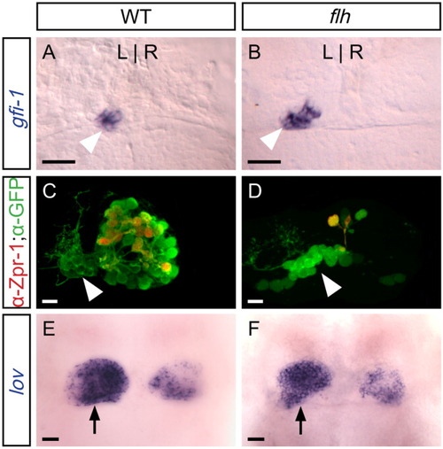
In flh mutants, parapineal specification is normal, but the pineal organ is reduced in size. All panels are dorsal views of the epithalamus of 4 d larvae. A: Expression of gfi-1 (blue), which is restricted to the parapineal organ (arrowhead), reveals ∼10 left-sided cells in WT and (B) flh larvae. C: In WT larvae, both the parapineal (arrowhead) and pineal express foxd3:gfp (green); many cells of the pineal organ are also labeled by the red-green cone marker Zpr-1 (Fret43) (red), but no parapineal cells are Zpr-1-labeled. D: In flh mutants, the pineal organ is drastically reduced in size, including fewer Zpr-1-labeled cells. As in WT, no Zpr-1-labeled cells are detected in the parapineal. E: At 4 d, lov expression is asymmetric in the habenular nuclei; the habenula adjacent to the parapineal (arrow) expresses lov extensively in WT and (F) flh mutants. Scale bars = 25 μm. |

Flh and Tbx2b do not regulate one another's expression. All panels are dorsal views of the epithalamus at the stage indicated. (A,B) tbx2b and (C,D) flh are expressed in the pineal complex anlage at the 6-somite and 24-hr stages. E,F: Double labeling for tbx2b (blue) and flh (red) reveals that they overlap in expression at the 6-somite and 24-hr stages; flh is expressed further medially than tbx2b at the 6-somite stage. G: In flh mutants, expression of tbx2b initiates on time and (H) is maintained, albeit in a reduced number of cells. I,J: In fby mutants (a lesion in tbx2b), expression of flh is unaltered relative to WT. Scale bars = 25 μm. EXPRESSION / LABELING:
|
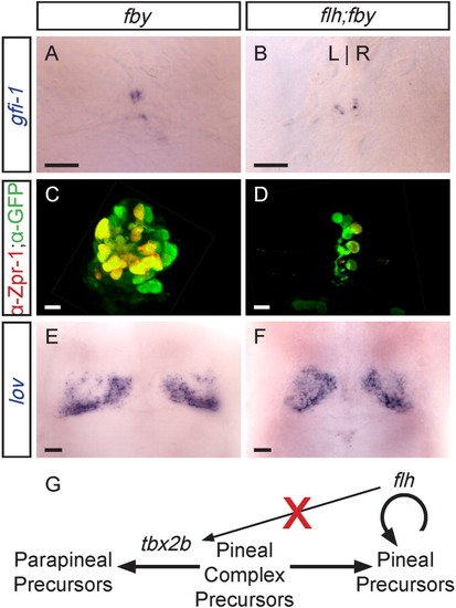
Double homozygous flh;fby mutants exhibit an additive phenotype. All panels are dorsal views of the epithalamus of 4 d larvae. A: In fby single mutants or (B) flh;fby double mutants, only a few disorganized parapineal cells are specified, and they fail to migrate to the left side of the brain. C: fby single mutants form a pineal organ. D: flh;fby double mutant larvae have both a reduced pineal, similar to flh single mutants (compare to Fig. 3D). E: In fby single mutants and (F) flh;fby double mutants, reduced lov expression is detected in the left habenula. Scale bars = 25 μm. G: Summary of tbx2b and flh activity in pineal complex development. The activity of tbx2b gives parapineal precursors their identity, while flh is needed for proliferation of pineal precursor cells. Expression of tbx2b does not require flh activity. |