|
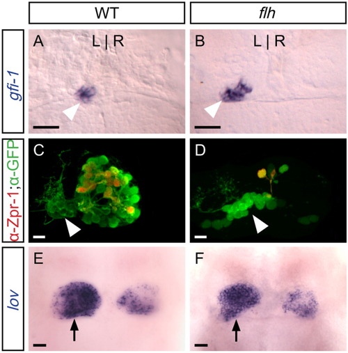
In flh mutants, parapineal specification is normal, but the pineal organ is reduced in size. All panels are dorsal views of the epithalamus of 4 d larvae. A: Expression of gfi-1 (blue), which is restricted to the parapineal organ (arrowhead), reveals ∼10 left-sided cells in WT and (B) flh larvae. C: In WT larvae, both the parapineal (arrowhead) and pineal express foxd3:gfp (green); many cells of the pineal organ are also labeled by the red-green cone marker Zpr-1 (Fret43) (red), but no parapineal cells are Zpr-1-labeled. D: In flh mutants, the pineal organ is drastically reduced in size, including fewer Zpr-1-labeled cells. As in WT, no Zpr-1-labeled cells are detected in the parapineal. E: At 4 d, lov expression is asymmetric in the habenular nuclei; the habenula adjacent to the parapineal (arrow) expresses lov extensively in WT and (F) flh mutants. Scale bars = 25 μm.
|