
- Title
-
Identification, tissue distribution and developmental expression of tjp1/zo-1, tjp2/zo-2 and tjp3/zo-3 in the zebrafish, Danio rerio
- Authors
- Kiener, T.K., Sleptsova-Friedrich, I., and Hunziker, W.
- Source
- Full text @ Gene Expr. Patterns

Zebrafish TJP expression patterns during early embryonic development. All figures depicting expression patterns show the embryos in the same orientation: anterior to the left, posterior to the right, dorsal to the top and ventral to the bottom. Expression of tjp1.1 (A,F,K), tjp1.2 (B,G,L), tjp2.1 (C,H,M), tjp2.2 (D,I,N) and tjp3 (E,J,O) was detected by whole mount in situ hybridization of embryos at the 64 cell stage (2 hpf) (A–E), at the sphere stage (4 hpf) (F–J) and at the shield stage (6 hpf) (K–O). Messenger RNA for all TJPs is maternally supplied (A–E). Hybridization signals remain strong after the midblastula transition, when embryonic transcription begins (F–J). Tjp2.1 mRNA is excluded from the nucleus (C, H, arrows), whereas tjp2.2 accumulates in the nucleus (D, I, arrows). At the stage of early gastrulation, TJP expression is weak but uniform (K–O). Tjp3 mRNA shows perinuclear localization (O, inset). |
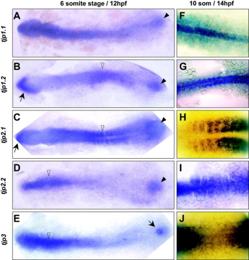
Zebrafish TJP expression patterns during early somitogenesis. Expression of tjp1.1 (A,F), tjp1.2 (B,G), tjp2.1 (C,H), tjp2.2 (D,I) and tjp3 (E,J) was detected by whole mount in situ hybridization of embryos at the 6 somite stage (12 hpf) (A–E) and 10 somite stage (14 hpf) (F–J). All TJP genes are expressed in the tail bud region (arrowheads) by 12 hpf (A–D). Tjp3 is expressed in a localized structure, most likely the Kupffer’s vesicle (E, arrow). The most anterior part of the embryo expresses tjp1.2 and tjp2.1 (B, C, arrows). The anterior axial mesoderm expresses lower levels of tjp1.2 (B) compared to other TJPs, whereas the anterior expression of tjp2.2 and tjp3 is restricted to this area (D, E, open arrowheads). In the posterior part of the embryo, the somitic mesoderm expresses high levels of tjp2.1 (C) and moderate levels of tjp1.2 (B, open arrowheads). TJP expression defines the identity of the posterior mesoderm. The axial mesoderm predominantly expresses tjp1.1 (notochord, F), and tjp1.2 (G) while somites express tjp2.1 (H), and tjp2.2 (I). Tjp3 is weakly detected in the posterior axial and paraxial mesoderm (J). EXPRESSION / LABELING:
|

Zebrafish TJP expression pattern during the end of somitogenesis. Expression of tjp1.1 (A,F,K), tjp1.2 (B,G,L), tjp2.1 (C,H,M), tjp2.2 (D,I,N) and tjp3 (E,J,O) was detected by whole mount in situ hybridization of embryos at the 18 somite stage (18 hpf) (A–E) and the Prim-5 stage (24 hpf) (F–O). Towards the end of somitogenesis, all TJP genes are expressed in six major domains, including the epidermis, somites, pronephric ducts (A–J) as well as the neural tube, eyes, and otic placodes (asterisk) (K–O). However, there is a selective expression pattern in the posterior part of the embryo, where tjp1.1 is expressed transiently in the hypochord (A, arrow), the blood island (A,F, arrowhead) and the notochord (A,F, open arrowhead). As observed during early somitogenesis, tjp1.2 is expressed both in the notochord (B, G, open arrowheads) as well as in the somites (B,G). Interestingly, the posterior part of the ventral spinal cord is transiently positive for tjp2.1 (C,H, arrowhead). Tjp3 expression is high in the pronephric ducts (E, arrow) and weak in the epidermis (E, arrowheads). EXPRESSION / LABELING:
|
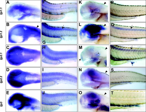
Zebrafish TJP expression pattern at the hatching and larval stages. Expression of tjp1.1 (A,F,K,P), tjp1.2 (B,G,L,Q), tjp2.1 (C,H,M,R), tjp2.2 (D,I,N,S) and tjp3 (E,J,O,T) was detected by in situ hybridization on whole mount embryos at the hatching stage (2 dpf) (A–J) and the larval stage (4 dpf) (K–T). Brain regions express high levels of all TJP genes (A–E). Note that tjp1.2 expression is predominant in the hindbrain (B, arrowhead). The epidermis, the somites and the pronephric ducts also express all five TJPs (F–J). The notochord retains tjp1.1 expression (F, open arrowhead). During the hatching period, the spinal cord starts to express tjp1.2 (G, arrow). The pectoral fin buds are tjp3 positive (E, arrowhead). At the beginning of the larval stage, the expression pattern of the five TJP genes converges. In the anterior part of the embryo, all five TJP genes are now expressed in the nose (arrow), otic capsule (asterisk), branchial arches (open arrowhead), and regions of the brain (K–O, see M). Tjp1.2 is the dominant gene expressed in the brain (L). As already observed at 2 dpf, tjp1.1 and tjp1.2 are expressed in the hindbrain (K,L, arrowheads), from where the other isoforms are absent (M,N,O, arrowheads). In the posterior part of the embryo, the pronephric ducts and intestine highly express TJP genes. Expression in the epidermis and somites is low and not readily detected by in situ hybridization (P–T), but the spinal cord now expresses high levels of both tjp1.2 and tjp2.1 (Q,R, arrows). |

Zebrafish TJP expression patterns in sensory anlagen. Expression of TJPs in sensory anlagen was detected by whole mount in situ hybridization. (A–C) Olfactory and otic placodes. Expression during sensory organ development is temporally regulated. Tjp3 is the first TJP family member to be expressed in a bell shaped area of the forehead (A, arrowhead), which will give rise to the olfactory placodes, and in the otic placodes (A, arrow) at 14 hpf. At 18 hpf, tjp3 is detected in paired domains of the olfactory placodes (B, arrowhead), the otic placodes (arrow), and the branchial arches vesicles (B, open arrowhead). Tjp2.1 is expressed in the otic placodes at 18 hpf (C, arrow) which is later than tjp3 but earlier than the other TJPs (see Fig. 4K–O). (D–I) Eyes. Tjp1.1 is the only family member expressed in the lens, starting at 1 dpf (D, arrow). By 4 dpf, TJP expression in the retina is mainly observed in the inner nuclear layer as well as in the ganglion cell layer. (E) tjp1.1 is expressed in the inner nuclear layer (arrowheads) and in the lens (arrows). (F) tjp1.2 is also expressed in the inner nuclear layer (arrowheads). In addition, there is a strong signal in the ganglion cell layer (open arrowheads). Tjp2.1 (G) and tjp2.2 (H) are found in the inner nuclear layer only (arrowheads). Tjp2.1 expression appears to be restricted to the outer part of the layer which contains bipolar and horizontal cells (G, arrow). (I) tjp3 is present in the nuclear layer (arrowhead) and also in the ganglion cell layer (open arrowhead). EXPRESSION / LABELING:
|
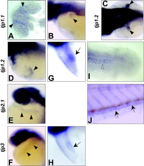
Zebrafish TJP expression patterns in other selected organs. Expression of TJPs in selected organs was detected by whole mount in situ hybridization. (A) Low expression of tjp1.1 is detected in the medial region of the eyes at 18 hpf (arrowheads). (B,C) Pectoral fin buds. The pectoral fin buds do not only express tjp3 (see Fig. 5E) but also tjp1.1 (A, arrowhead) and tjp1.2 (B, arrowheads) at 2–3 dpf. (D–F) Duct of Cuvier. The Duct of Cuvier expresses tjp1.2 (D), tjp2.1 (E), and tjp3 (F, arrowheads). (G,H) Heart. The newly forming heart transiently expresses high levels of the tjp1.2 (G, arrow) and low levels of tjp3 (H, arrow) at 2 dpf. (I) Notochord. The posterior part of the notochord retains tjp1.2 expression up to 3 dpf (arrowhead). (J) Caudal vein. The caudal vein expresses tjp2.1 at 3 dpf (arrows). EXPRESSION / LABELING:
|
Reprinted from Gene expression patterns : GEP, 7(7), Kiener, T.K., Sleptsova-Friedrich, I., and Hunziker, W., Identification, tissue distribution and developmental expression of tjp1/zo-1, tjp2/zo-2 and tjp3/zo-3 in the zebrafish, Danio rerio, 767-776, Copyright (2007) with permission from Elsevier. Full text @ Gene Expr. Patterns