
Fig. 3
|
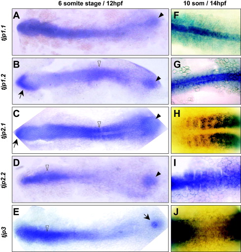
Fig. 3 Zebrafish TJP expression patterns during early somitogenesis. Expression of tjp1.1 (A,F), tjp1.2 (B,G), tjp2.1 (C,H), tjp2.2 (D,I) and tjp3 (E,J) was detected by whole mount in situ hybridization of embryos at the 6 somite stage (12 hpf) (A–E) and 10 somite stage (14 hpf) (F–J). All TJP genes are expressed in the tail bud region (arrowheads) by 12 hpf (A–D). Tjp3 is expressed in a localized structure, most likely the Kupffer’s vesicle (E, arrow). The most anterior part of the embryo expresses tjp1.2 and tjp2.1 (B, C, arrows). The anterior axial mesoderm expresses lower levels of tjp1.2 (B) compared to other TJPs, whereas the anterior expression of tjp2.2 and tjp3 is restricted to this area (D, E, open arrowheads). In the posterior part of the embryo, the somitic mesoderm expresses high levels of tjp2.1 (C) and moderate levels of tjp1.2 (B, open arrowheads). TJP expression defines the identity of the posterior mesoderm. The axial mesoderm predominantly expresses tjp1.1 (notochord, F), and tjp1.2 (G) while somites express tjp2.1 (H), and tjp2.2 (I). Tjp3 is weakly detected in the posterior axial and paraxial mesoderm (J).
Reprinted from Gene expression patterns : GEP, 7(7), Kiener, T.K., Sleptsova-Friedrich, I., and Hunziker, W., Identification, tissue distribution and developmental expression of tjp1/zo-1, tjp2/zo-2 and tjp3/zo-3 in the zebrafish, Danio rerio, 767-776, Copyright (2007) with permission from Elsevier. Full text @ Gene Expr. Patterns