
- Title
-
The activity of Neurogenin1 is controlled by local cues in the zebrafish embryo
- Authors
- Blader, P., Fischer, N., Gradwohl, G., Guillemot, F., and Strähle, U.
- Source
- Full text @ Development
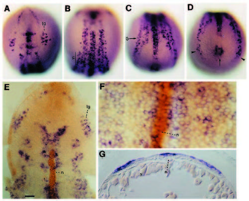
ngn1 is expressed broadly in distinct domains in the neural plate. (A-D) Dorsal views of an embryo at the 3-somite stage showing ngn1 expression. Orientation is anterior up; (A) view onto anterior neural plate; (B) view onto neural plate at the posterior hindbrain/anterior trunk level; (C) view onto neural plate at trunk level; (D) view onto tail bud. Expression is detected in the center (arrow) and lateral aspects of the tailbud (arrowheads). (E,F) Flat preparations of 3-somite embryos showing ngn1 expression (purple) and anti-Notail antibody (brown) marking the nuclei of the notochord (n). E shows a flat preparation of the anterior neural plate while F shows an area at the trunk level as indicated by line, F in B. View onto neural plate, anterior up. (G) Transverse section through 3-somite stage embryo at the level indicated by arrow marked G in C. Dorsal is up. Abbreviations: tg, trigeminal ganglion; n, notochord; y, yolk. Scale bar represents 50 μm in A-E; 12 μm in F and G. EXPRESSION / LABELING:
|

ngn1 is expressed earlier and more widely than zebrafish nrd and isl-1. (A,B) nrd expression in 1-somite stage embryos. nrd is expressed in a row of cells in the anterior neural plate (arrowhead in A), the trigeminal ganglion (tg) and in two lateral rows of cells in the trunk neural plate (arrow in B). Dorsal views onto anterior neural plate (A) and at mid trunk level (B). (C,D) isl-1 expression in 1s embryos. isl-1 is expressed in the pillow (arrow head in C); the trigeminal ganglion (tg) and in two medial and two lateral rows of cells in the posterior neural plate (arrows in D). Dorsal views onto anterior neural plate (C) and at mid trunk level (D). (E) ngn1 expression in 1-somite stage embryo. Dorsal view onto neural plate at posterior hindbrain/anterior trunk level. (F) ngn1 expression at the 90% epiboly stage; view is dorsal onto the hindbrain anlage, anterior up. ngn1 is already expressed whereas isl-1 and nrd are not expressed in the late gastrula. The lateral extent of the neural plate is marked by arrowheads; arrows indicate the trigeminal ganglia, which are widely spaced at this stage. Abbreviations: tg, trigeminal ganglion. EXPRESSION / LABELING:
|
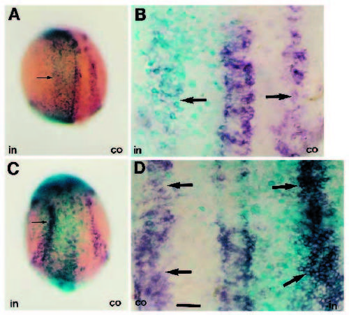
ngn1 expression is regulated by Delta. (A,B) Embryos at the 3- somite stage co-injected with X-Delta-1 and β-gal RNA into one blastomere at the 2-cell stage. Embryos were stained for β-gal activity (turquoise) before in situ hybridisation with ngn1 antisense probe (purple). ngn1 expression (arrows) is strongly reduced or completely abolished in the injected side (turquoise). (C,D) Embryos at the 3- somite stage co-injected with the antimorphic X-Delta-1Stu and bgal RNA into one blastomere at the 2-cell stage. Expression of X-Delta- 1Stu results in up-regulation of ngn1 expression (purple, arrows) in the injected side (turquoise) in comparison to the uninjected control side. (A,C) Whole mounts in dorsal views onto the neural plate at the level of posterior hindbrain/anterior trunk; anterior up. B and D show flat preparation of different embryos from those in A and C, respectively; anterior up. Abbreviations: in, injected side; co, uninjected control side. Scale bar, 60 μm in A,C and 25 μm in B, D. |
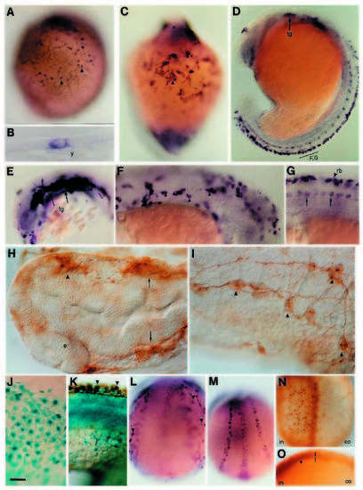
Misexpression of ngn1 induces ectopic neurons. (A) Ventral view of 3-somite stage, ngn1 RNA-injected embryo showing ectopic isl-1 expression in the non-neural ectoderm of the yolk sac (arrowheads). (B) Transverse section through yolk sac of ngn1 RNA-injected embryo showing ectopic isl-1-expressing cell in the yolk sac ectoderm. (C,D) ngn1 RNA-injected embryo (C), and uninjected control (D), at the 18- somite stage hybridised to isl-1 probe. Ventral view of injected embryo (C) shows the ectopic isl-1-expressing cells in the yolk sac (arrowhead) forming cell clusters at this stage. Control embryo (D) is shown in lateral view. (E-G) ngn1-injected embryos analysed for isl-1 expression at the 18-somite stage. (E) Lateral view of the head with an expanded trigeminal ganglion (arrows, tg; compare to D); (F,G; lateral views, dorsal up). Optical sections at the level of the surface ectoderm (F) and the midline of the neural tube of the same injected embryo. Position of frames along the anteroposterior axis is indicated in D. While injected embryos develop numerous isl-1- expressing cells in the surface ectoderm (arrowheads in F) the number of isl-1-expressing Rohon- Beard (rb in G) and motor neurons (arrow in G) in the neural tube is unaltered in injected embryos (compare with uninjected control in D). (H) Head of ngn1 RNA-injected 24h embryo immunohistochemically stained with antibody zn12 which recognises the L2/HNK1 epitope. Embryo (dorsal view onto head, anterior left) shows loss of an eye and concomitant unilateral ectopic formation of a ganglion-like structure (arrowhead) and unilateral expansion of the trigeminal ganglion (arrows). (I) Optical section through surface ectoderm at the level of the hindgut of ngn1 RNA-injected 24h embryo immunohistochemically stained with antibody zn12. Neurons (arrowheads) with processes which contact each other develop ectopically in the epidermis of injected embryos. (J, K) View onto yolk sac (J) and optical section through body axis (K) of embryos injected with β-gal RNA. Embryos were stained for β-gal activity (turquoise) and immunohistochemically with antibody zn12 (brown). Expression of β-gal did not cause the formation of ectopic neurons. Only the normal expression of the L2/HNK1 epitope could be detected in injected embryos (K, arrowheads). (L,M) ngn1-myc RNAinjected embryo (L) and uninjected control (M) at the 3-somite stage hybridised to isl-1 probe. The embryo shown in L was injected in both blastomeres at the two-cell stage. Ectopic isl-1-expressing cells are found lateral to, but not within the neural plate. Views onto the neural plate at the level of the posterior hind brain/anterior trunk, anterior up. The optical section through the embryo shown in L is taken from a deeper position than the section in M to show ectopic neurons (arrowheads) on both sides of the neural plate. The normal pattern of isl-1 expression in the neural plate is therefore out of focus in the injected embryo. (N,O) Embryos injected with ngn1-myc RNA in one blastomere at the two-cell stage and immunohistochemically stained with anti-myc antibody 9E10 at the 3-somite stage to visualise ectopic expression of Ngn1-MYC. View is dorsal with anterior up (N) and optical transverse section through neural plate with dorsal up (O). Ngn1-MYC is strongly expressed in the neural plate (arrowhead in O) in the injected side (in). Lack of immunostaining on the uninjected side (co) shows that the MYC immunoreactivity is specific. Abbreviations: e, eye; y, yolk; tg, trigeminal ganglion; rb; Rohon-Beard sensory neurons; in, injected side of embryo; co, control side of embryo. Scale bar represents 50 μm in A,C,D,L,M,N,O; 25 μm in E,F,GJ,K; and 12 μm in B,I. |

Misexpression of ngn1 causes ectopic development of distinct types of neurons (arrowheads) in a region specific manner. (A,B) ngn1- myc-injected (A) and uninjected control (B) embryo hybridised to isl-1 probe at the 3-somite stage. Lateral views; anterior up. (C,D) ngn1-mycinjected (C) and uninjected control (D) showing isl-2 expression at the 24h stage. Ectopic isl-2 expression is detectable in the non-neural ectoderm. Optical sections through surface ectoderm at the level of the hindgut extension. Dorsal up, anterior left. (E,F) Ectopic nrd expression in ngn1-myc-injected (E) and uninjected (F) control embryo at the 3-somite stage. nrd-expressing cells are detectable in the anterior half of the yolk sac ectoderm. Lateral views, anterior up. (G-I) Ectopic nrd expression is detectable over the yolk (G) and over the surface ectoderm covering the body axis in 24h ngn1-myc-injected (I) but not in uninjected controls (H). nrd expression in the nasal placodes is expanded in ngn1-myc-injected embryos (arrow). Ventral views, anterior up (G,H). Lateral view, anterior left, dorsal up (I). (J-L) Ectopic zash1b expression is found in cells lateral to the body axis in ngn1-myc-injected (J,L) but not in uninjected controls (K). Dorsal view, anterior up (J,K). Lateral view, dorsal up, anterior left (L). (M) ngn1-myc-injected embryos express hlx-1 ectopically in cells in the endoderm. Scale bar: 50 μm (A,B,E-H,J,K), 25 μm (C,D,I,L) and 12 μm (M). |

Co-injection of ngn1 and RNA for a dominant negative regulatory subunit of mouse protein kinase A (dnReg) leads to formation of ectopic lim-3-expressing cells. (A) Embryo injected with ngn1 and dnReg RNA. Arrows indicate ectopic lim-3 expression. (B) Embryo injected with dnReg RNA alone. Arrowheads indicate dorsal expansion of lim-3 expression. (C) Uninjected control embryo stained with lim-3 RNA. Embryos are 24 hours old and are shown anterior left and dorsal up. The embryo in A is slightly tilted to show ectopic lim-3-expressing cells in the ectoderm and lim-3 expression in the neural tube in one optical plane. Scale bar: 25 μm (A) and 12 μm (B,C). |
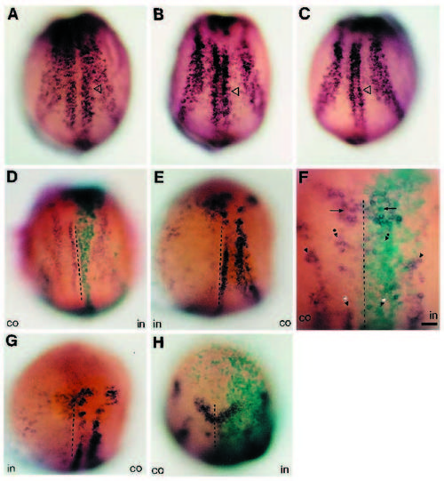
Expression of ngn1 is modulated by Shh. (A-C) ngn1 expression in 3-somite stage embryo injected in both blastomeres at the 2-cell stage with dnReg (A), shh RNA (B) and an uninjected control (C). The medial rows of ngn1 expression in the neural plate appear broadened in dnReg- and shh-injected embryos (open arrowhead). (D-G) Embryos at the 3-somite stage injected with a constitutively active PKA (PKA*) and β-gal RNA; only one blastomere of the 2- cell stage embryo was injected. Embryos were stained for ngn1 expression (purple) and β-gal enzymatic activity (turquoise). Control and injected sides are indicated by co and in, respectively. Arrowheads in F indicate dorsal neuron precursors (alone), motor neuron precursors (with asterisks) and a bilateral pair of hindbrain nuclei (with dots); arrows in F mark the midbrain/hindbrain boundary. The midline of the neural plate is indicated by a dotted line. (H) Embryo injected with β-gal RNA alone. Dorsal views; anterior up. |