
- Title
-
Translation repression by maternal RNA binding protein zar1 is essential for early oogenesis in zebrafish
- Authors
- Miao, L., Yuan, Y., Cheng, F., Fang, J., Zhou, F., Ma, W., Jiang, Y., Huang, X., Wang, Y., Shan, L., Chen, D., Zhang, J.
- Source
- Full text @ Development
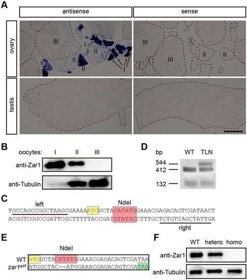
Generation of a zar1 mutant with TALENs in zebrafish. (A) In situ hybridization on cryosections of wild-type gonads with zar1 antisense probe and sense probe. Scale bar: 200 μm. (B) Immunoblotting to detect Zar1 protein in stage I, stage II and stage III oocytes. Ten oocytes were lysed for each stage. (C) The TALEN sequences for zar1 mutant generation. (D) Digestion of PCR products from wild-type (WT) and zar1 TALEN mRNA injected embryos (TLN) with NdeI restriction enzyme. (E) DNA sequences of the zar1gd5 mutant fish line. A premature stop codon was generated. (F) Western blot of Zar1 in gonads from WT, heterozygotes (hetero) and homozygotes (homo) at 25 dpf. DNA sequences highlighted in yellow are the start codon; red, NdeI recognition sites; green, premature stop codon. EXPRESSION / LABELING:
PHENOTYPE:
|

Loss of Zar1 causes all-male phenotype in zebrafish. (A) Analysis of genders of zar1 homozygotes (homo), heterozygotes (hetero) and wild-type siblings. (B-D) Histological analysis of gonads of zar1 homozygotes and sibling controls by H&E staining. About half of the gonads (20/37) from wild-type and zar1 heterozygotes were ovaries (B) while the other half (17/37) were testes (C). All the 13 gonads from zar1 homozygotes were testes (D). (E) Schematic diagram of the Tg(zp3b:zar1) transgenic construct. egfp coding sequences were placed under control of the cmlc2 promoter to help visually identify transgenic fish. (F) Gender analysis of zar1−/− homozygotes with or without EGFP signal, indicating Tg(zp3b:zar1) transgene. Females were recovered only from zar1−/− homozygotes with the Tg(zp3b:zar1) transgene. (G,H) H&E staining of ovaries of zar1+/− heterozygotes and zar1−/− homozygotes on the Tg(zp3b:zar1) transgenic backgrounds. Ovaries from zar1−/− homozygotes rescued with the Tg(zp3b:zar1) transgene are normal histologically. Tg(zp3b:zar1), Tg(zp3b:zar1,cmlc2:EGFP); sg, spermatogonia; sc, spermatocytes; sp, sperm; I,II,III, oocyte stage I, II or III. Scale bars: 40 μm. PHENOTYPE:
|
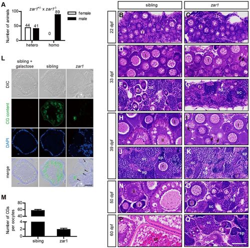
Zar1 deficiency causing all-male phenotype is due to female-to-male sex reversal. (A) Analysis of genders of zar1 homozygotes (homo) and heterozygous siblings (hetero). zar1+/− heterozygous females were crossed with zar1−/− homozygous males and genders of their progenies were analyzed. (B-K,N-Q) Gonad development of zar1 homozygotes and control siblings analyzed by H&E staining. At 22 dpf, zebrafish gonads are undifferentiated. Gonads of zar1 homozygotes and control siblings are indistinguishable histologically (B,C). At 33 dpf, WT gonads differentiate into immature ovaries and immature testes. Minor developmental abnormalities are observed in zar1 homozygotes. Oocytes in zar1 homozygotes are similar to those in WT in size and morphology, but aberrant vesicles (arrow indicated) are observed in ooplasm of a few zar1 homozygous oocytes (D,E). Testis development in zar1 homozygotes is normal compared with that in control siblings (F,G). At 39 dpf, ovarian developmental abnormality in zar1 homozygotes becomes more pronounced (H,I), while testis development in zar1 homozygotes is similar to the controls (J,K). Immature ovaries in control siblings contain stage I and stage II oocytes (H) while oocytes in zar1 homozygotes resemble stage I oocytes, indicating oogenesis arrest in zar1 homozygotes. In addition, aberrant vesicles (arrow) in zar1 homozygotes increase significantly in size and number. (L) MPA (Maclura pomifera agglutinin) staining of ovary sections. Juveniles at 37-40 dpf fixed with 4% PFA were embedded in paraffin and sections stained with MPA; 0.5 M D-galactose inhibited MPA staining. MPA specifically stains CGs in control siblings. The aberrant vesicles in zar1 mutant ovaries are MPA positive (arrows). (M) Comparison of CG numbers (mean±s.e.m.) between zar1 mutants (n=11) and siblings (n=11). At 50 dpf, stage II oocytes are observed in heterozygous and wild-type ovaries (N), but large numbers of oocytes underwent atresia in zar1 homozygotes, and spermatogonia and spermatocytes appear among oocytes, indicating transitional ovaries (ovotestis) (O). At 60 dpf, stage III oocytes were seen in the control ovaries (P); in contrast, very few oocytes remain in ovotestis of zar1 mutants and spermatogenesis has progessed further (Q). At 22 dpf, six juveniles were analyzed for each genotype. At 33 dpf, 39 dpf, 50 dpf and 60 dpf, 20 juveniles per stage were analyzed for each genotype. Stars indicate degenerating oocytes. sg, spermatogonia; sc, spermatocytes; sp, sperm. Scale bar: 40 μm. PHENOTYPE:
|
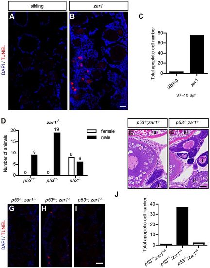
Zar1 deficiency causing all-male phenotype is due to p53-mediated apoptosis. (A-C) TUNEL staining of ovary sections of zar1 homozygotes and control siblings at 37-40 dpf. Obvious apoptotic cells are observed in immature ovaries in zar1 homozygotes (A), but not in sibling controls (B). Quantification of apoptotic cells is shown in C; 18 sections from 6 juveniles were counted for each genotype. (D) Gender analysis of zar1−/− homozygotes on different p53 genotype backgrounds (p53+/+, p53+/− and p53−/−). Females are only observed in p53−/−;zar1−/− double homozygous mutants. (E,F) H&E staining of sections of p53−/−;zar1+/− and p53−/−;zar1−/− adult ovaries. p53−/−;zar1−/− ovaries are morphologically normal. (G-J) TUNEL staining of ovary sections of p53−/−;zar1−/− ovaries at 37-40 dpf with p53−/−;zar1+/− and p53+/−;zar1−/− ovaries as controls. (J) Quantification of apoptotic cells. Nine sections from three juveniles were counted for each genotype. Scale bar: 20 μm. PHENOTYPE:
|

EE2 treatment restores ovarian development in zar1 homozygous mutant. (A) Statistics for sex ratio of zar1 homozygous and heterozygous mutants after EE2 treatment. (B,C) H&E staining of ovarian tissue in EE2-treated zar1 homozygotes and heterozygotes. Similar to zar1 heterozygous ovaries, the treated homozygous mutant ovaries possess oocytes at all stages. (D) btg2 and traf4a expression analyzed by qPCR in zar1 homozygotes (n=6) and heterozygotes (n=6). (E) atf3 expression analyzed by qPCR in zar1 homozygotes (n=7) and heterozygotes (n=8) with or without EE2 treatment. Gene expression was normalized to expression of elongation factor 1 alpha (ef1a). hetero, zar1 heterozygotes; homo, zar1 homozygotes; EE2-hetero, EE2-treated zar1 heterozygotes (n=6); EE2-homo, EE2 treated zar1 homozygotes (n=6). Data are mean±s.e.m. **P<0.01, ***P<0.001; ns, not significant. Scale bar: 20 μm. EXPRESSION / LABELING:
PHENOTYPE:
|

ZFIN is incorporating published figure images and captions as part of an ongoing project. Figures from some publications have not yet been curated, or are not available for display because of copyright restrictions. EXPRESSION / LABELING:
PHENOTYPE:
|
|
ZFIN is incorporating published figure images and captions as part of an ongoing project. Figures from some publications have not yet been curated, or are not available for display because of copyright restrictions. EXPRESSION / LABELING:
PHENOTYPE:
|
|
ZFIN is incorporating published figure images and captions as part of an ongoing project. Figures from some publications have not yet been curated, or are not available for display because of copyright restrictions. EXPRESSION / LABELING:
PHENOTYPE:
|
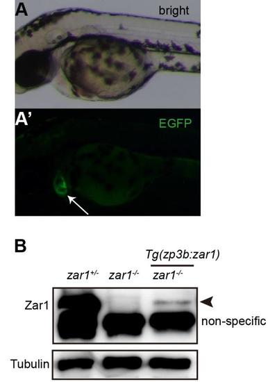
Zar1 is expressed in zar1 homozygous females with the Tg(zp3b:zar1,cmlc2:EGFP) transgene.(A, A') Heart specific EGPF signal (arrow) seen in transgenic embryos. (B) Western blot to detect Zar1 protein in adult ovaries. Zar1 was expressed in the transgene rescued adult zar1 homozygous ovaries (arrowhead). Compared with Zar1 expression level in zar1 heterozygous mutant ovaries (hetero), Zar1 expression level in transgene rescued zar1 homozygous (homo) mutant ovaries was low. The smaller bands are non-specific. |
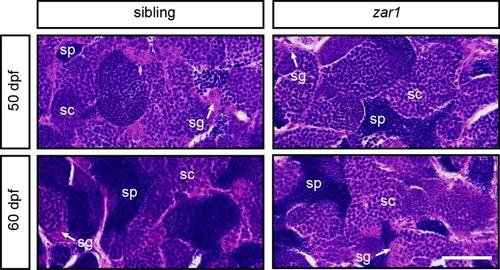
Spermatogenesis is not affected in zar1 mutant males.Histology analysis of testes of zar1 homozygous males and control siblings at 50 dpf and 60 dpf. Spermatogenesis in zar1 homozygous males resembles that in control siblings. sg: spermatogonia; sc: spermatocytes; sp: sperm; Scale bar: 0.04 mm. |
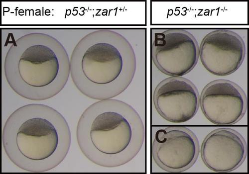
The chorions of eggs from p53-/-;zar1-/- failed to elevate normally.Five p53-/-;zar1+/- females and five p53-/-;zar1-/- females were crossed with wild-type males. The chorions of eggs from p53-/-;zar1+/- lifted normally (A). The chorions of eggs from p53-/-;zar1-/- failed to lift. Cleaved embryos (B) and uncleaved embryos (C); P-female: genotypes of the mothers |

The chorions of zar1 mutant eggs failed to elevate normally.EE2 treated females were crossed with wild-type males. Embryos from 6 heterozygous females (EE2-hetero) and 6 homozygous females (EE2-homo) were analyzed. The chorions of zar1 homozygotes failed to lift properly compared with those of zar1 heterozygotes. P-female: genotypes of the mothers. |