
Fig. 2
|
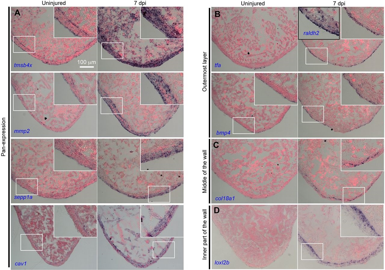
Fig. 2
New markers of epicardial cells. In situ hybridization for selected genes on sections from regenerating ventricles at 7 days post injury (dpi) in cmlc2:CreER; bactin2:loxP-mCherry-STOP-loxP-DTA animals treated with tamoxifen, as compared with ventricles from bactin2:loxP-mCherry-STOP-loxP-DTA animals treated with tamoxifen (uninjured). raldh2 served as a 7dpi marker for endocardium and the outermost layer of epicardium. The selected genes show: (A) pan-epicardial expression; (B) exclusive expression in the outermost layer of the ventricle wall; (C) expression within the wall; and (D) expression in the inner part of the wall but absent from the outermost layer. Insets are high-magnification views of the boxed areas. All of the genes were tested on sections from at least four uninjured hearts and five injured hearts. Each heart was represented by at least six sections. When similar patterns were detected in most of the six sections of each heart, and in all of the examined hearts, a conclusion of positive expression was made.