
Fig. 3
|
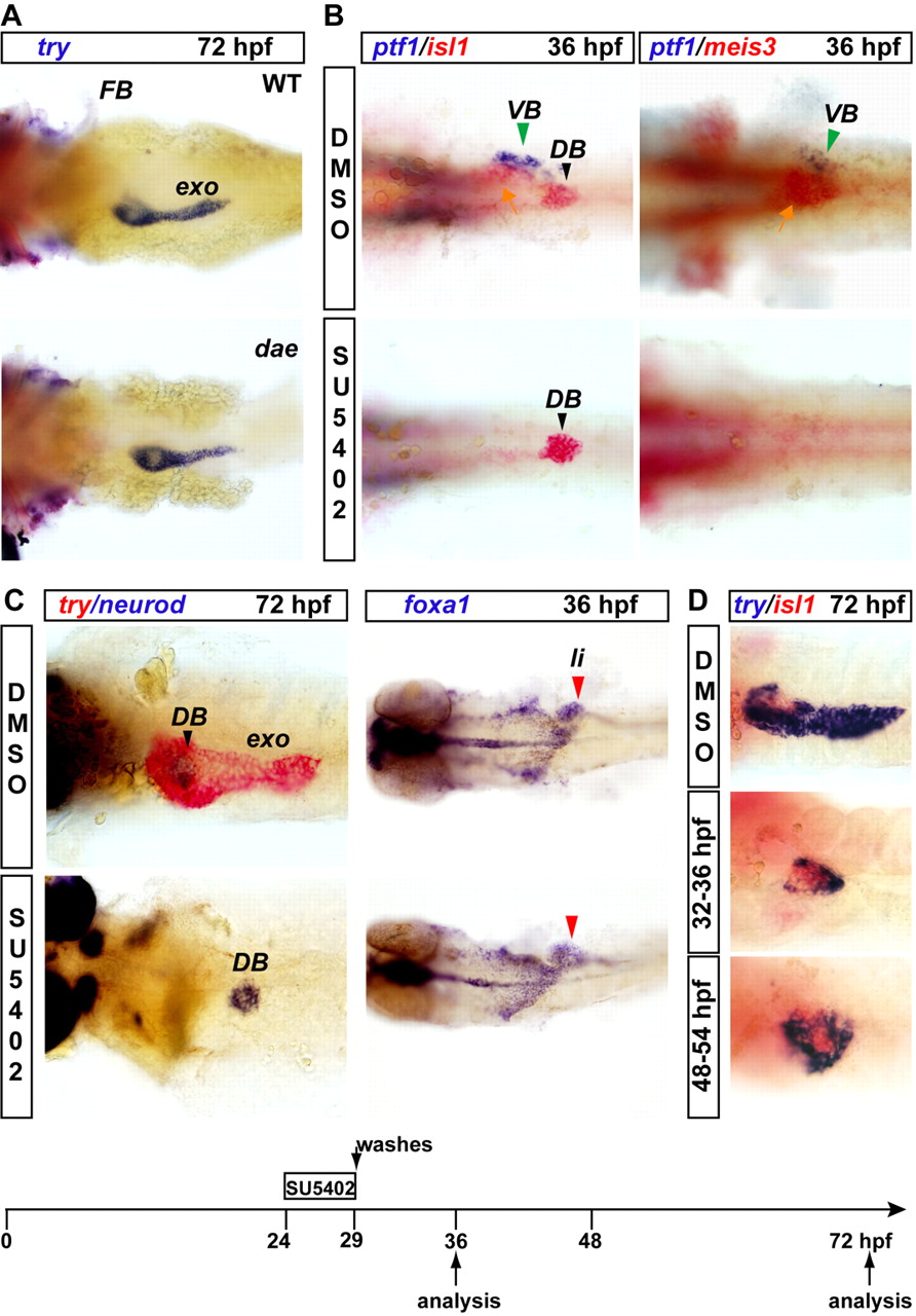
Fig. 3 Inhibition of FGF signaling, but not Fgf10, impairs specification of the ventral pancreatic bud and the expression of isl1 and meis3 in the adjacent pancreatic LPM. (A) trypsin (try) expression analysis in wild-type embryos (WT, top) and in fgf10-/- mutants (dae, bottom) at 72 hpf. Note the underdeveloped pectoral fin bud in the dae mutant. (B) Expression of ptf1a (blue), isl1 and meis3 (red) at 36 hpf in embryos treated with the FGF signaling inhibitor SU5402 from 24 to 29 hpf. (C) Expression of trypsin (red) and neurod (blue) at 72 hpf after the same treatment as in B and analysis of the endodermal marker foxa1 at 36 hpf upon SU5402 treatment. Note that the liver and the rest of the endoderm are clearly labeled whereas, at this stage, the pancreas is almost undetectable. The SU5402 treatment analyzed in B and C is schematized at the bottom of the panel. (D) trypsin (blue) and isl1 (red) expression at 72 hpf in embryos exposed to SU5402 from 32 to 36 hpf and from 48 to 54 hpf. The green arrowhead indicates the ventral pancreatic bud (VB); the black arrowhead indicates the dorsal bud (DB); and the yellow arrow indicates the pancreatic LPM adjacent to the ventral bud. exo, exocrine tissue; li, liver; FB, pectoral fin bud.