
Fig. 4
|
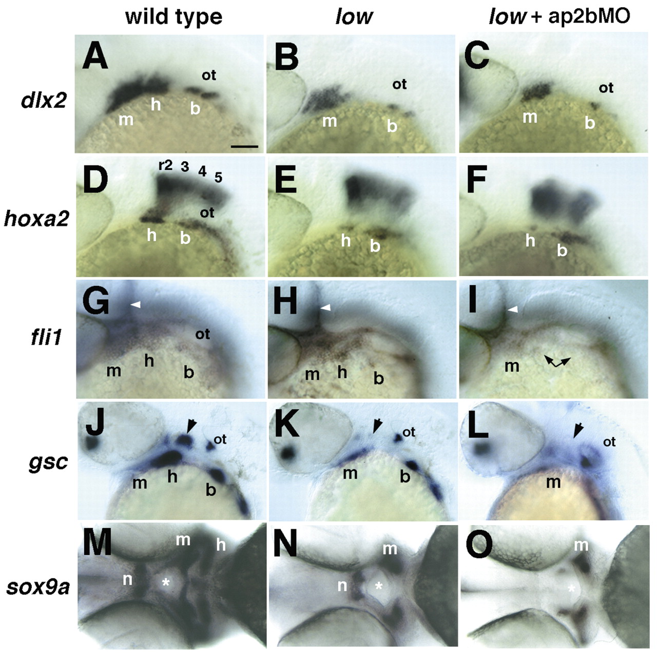
Fig. 4 Defects in pharyngeal arch development in low mutants and in larvae injected with ap2bMO. Whole-mount in situ hybridization with riboprobes to dlx2 (A-C; 28 hpf), hoxa2 (D-F; 28 hpf), fli1 (G-I; 28 hpf), gsc (J-L; 40 hpf) and sox9a (M-O; 52 hpf). (A-L) All embryos are shown in lateral view, except M-O, which are ventral views. Wild-type (A,D,G,J,M), low (B,E,H,K,N) and injected low mutants (C,F,I,L,O). Arrowheads in G-I indicate vascular expression of fli1. Arrows in J-L indicate the hyosymplectic condensation in the dorsal second arch. Asterisks in M-O indicate the mouth. All five NCC markers are reduced in the second pharyngeal arch in low mutants, but only fli1, gsc and sox9a defects are enhanced by Tfap2b depletion (L,P,T). b, branchial arches; h, hyoid arch; hb, hindbrain; m, mandibular arch; n, notochord. Scale bars: 100 µm.