
Fig. 5
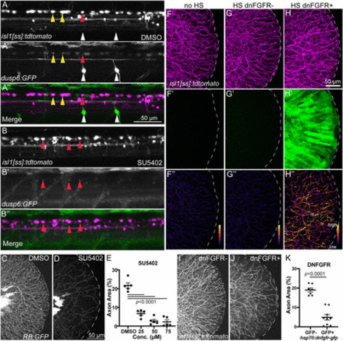
Inhibition of Fgf signaling induces loss of established sensory axons. A,B, Lateral views of isl1[ss]:tdTomato; dusp6:eGFP dorsal neurons in DMSO- (A) and SU5402-treated (B) 4 dpf larvae. Many RB neurons and motor neurons are GFP+ in DMSO-treated larvae but SU5402 treatment leads to loss of almost all GFP signal in dorsal neurons. Yellow arrowheads = GFP+ RB neurons, red arrowheads = GFP− RB neurons, white arrowheads = GFP+ motor neurons. C,D, Lateral views of RB:GFP caudal tails at 5 dpf treated for 48 h with 50 μM SU5402 (D), an Fgfr inhibitor, displayed substantial loss of RB axons in the tail compared to DMSO controls (C). E, Quantification of the dose-dependent effect of SU5402 on tail axon density. DMSO = 21.7 ± 1.5%, 25 μM = 6.7 ± 0.9%, 50 μM = 2.7 ± 0.9%, 75 μM = 2.3 ± 1.1%, analyzed by one-way ANOVA with post hoc Tukey's test (F = 68.46, p < 0.0001). F–H, Lateral views of caudal tails in isl1[ss]:tdTomato larvae following 30 min heat-shock. (F’–H’) EGFP channel. (F’’–H’’) Masked images showing axon-only EGFP expression. Compared to larvae without the hsp70:dn-fgfr1-EGFP transgene (F) or larvae with the dn-FGFR1 transgene that were not heat-shocked (G), hsp70:dn-fgfr1-EGFP+ larvae that underwent heatshock displayed detectable EGFP signal both throughout the tail tissue (H’) and specifically in isl1[ss]:tdTomato-labeled axons (H’’). I,J, Lateral view of isl1[ss]:tdTomato+ control (I) and isl1[ss]:tdTomato; hsp70:dn-fgfr1-GFP+ (J) caudal tails heatshocked at 4 dpf and imaged at 5 dpf. Compared to control (I), hsp70:dn-fgfr1-GFP+ larvae (J) had a reduction in RB axons similar to SU5402 treatment. K, Quantification of tail axon-density following heat-shock. Dnfgfr1-gfp− = 19.0 ± 1.0%, dnfgfr1-fgp+ = 4.9 ± 1.4%, analyzed by t test. |