
Fig. 4
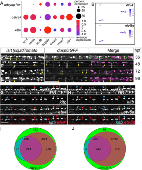
RBs express canonical downstream Fgf-signaling targets. A, Dot plot shows expression of canonical Fgf targets in all three RB clusters. B, Feature plots of two Fgf targets, etv4 and etv5a, show expression in kitb+ and calca+ clusters from the UMAP depicted in Figure 1F. C–F, Lateral views of dorsal neurons of live isl1[ss]:tdTomato; dusp6:GFP larvae at 36, 48, 72 and 96 hpf. Note that many RBs are GFP+ indicating active canonical Fgf signaling at these stages. G,H, Lateral views of FISH staining in RB:GFP larvae at 72 hpf for expression of kitb (cyan arrowheads) and downstream Fgf signaling targets (G) etv4 (red arrowheads) or (H) etv5a (red arrowheads). Yellow arrowheads = dusp6+ RBs, Red arrowheads = dusp6− RBs, scale bars = 50 μm. I,J, Summed counts of RB markers in larvae with respective sets of FISH probes visualized in (G,H) (N = 690 and 551, respectively). The majority of RBs express etv4 or etv5a and nearly all kitb+ RBs express etv4/etv5a. |