
Fig. 4
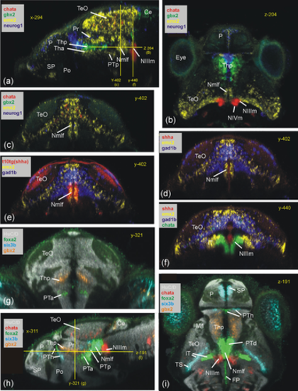
Basal prosomere P1 zebrafish gene expression (region of the Nmlf). (a–c) Region of the Nmlf in three atlas planes (a-c: emx2). Note that emx2, in addition to alar and basal P1 domains, has a thalamic domain (Tha; see text). In addition, gbx2/neurog1 expression shows the thalamus (Thp; see text) and chata marks midbrain oculomotor/trochlear nuclei as reference points. (d–f) Transverse sections show some differences between basal P1 and basal midbrain using emx2, gad1b, and shha expression. (g–i) Three atlas planes explain the position of basal P1 within basal diencephalon using basal plate marker gene foxa2. Ce, cerebellum; FP, (hindbrain) floor plate; lT, lateral midbrain tegmentum; M1, early migrated pretectal area; Nmlf, region of the nucleus of the medial longitudinal fascicle; NIIIm, oculomotor nucleus; NIVm, trochlear motor nucleus; P, pallium; Pi, pineal; Po, preoptic area; Pr, pretectum; PTa, anterior part of posterior tuberculum; PTh, prethalamus; PTp, posterior part of posterior tuberculum; SP, subpallium; TeO, tectum opticum; Tha, anterior thalamus; Thp, posterior thalamus; TS, torus semicircularis; ZLI, zona limitans intrathalamica. |