FIGURE

FIGURE 5

ID
ZDB-FIG-240323-31
Publication
Wang et al., 2024 - Deficiency of heme oxygenase 1a causes detrimental effects on cardiac function
Other Figures
All Figure Page
Back to All Figure Page
FIGURE 5

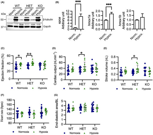
Hypoxia enhances cardiac output in HET(52del) larvae. (A) Representative immunoblots of β‐tubulin and Gapdh in WT, HET(52del), and KO(52del) at normoxic and hypoxic (3% O2 for 24 h) conditions. Gapdh serves as an internal control. Norm. normoxia; Hypox. Hypoxia. (B) RT‐qPCR analyses indicate that hypoxia induces upregulation of epoa and hmox1a, but not hmox1b, in WT larvae. (C–G) Hypoxia enhances ejection fraction (C) in WT and HET(52del) larvae, and cardiac output (D) and stroke volume (E) in HET(52del) larvae, but not in KO(52del), compared to normoxia. Hypoxia has no effect on heart rate (F) or diastolic area (G) in the three genotypic groups. WT, normoxia n = 15, hypoxia n = 9; HET(52del), normoxia n = 19, hypoxia n = 10; KO(52del), normoxia n = 16, hypoxia n = 10. Data are presented as mean ± SD. Two‐sample t‐test. *p < 0.05, **p < 0.01, ***p < 0.001.

Expression Data

Expression Detail
Antibody Labeling
Phenotype Data

Phenotype Detail
Acknowledgments
This image is the copyrighted work of the attributed author or publisher, and ZFIN has permission only to display this image to its users. Additional permissions should be obtained from the applicable author or publisher of the image. Full text @ J. Cell. Mol. Med.