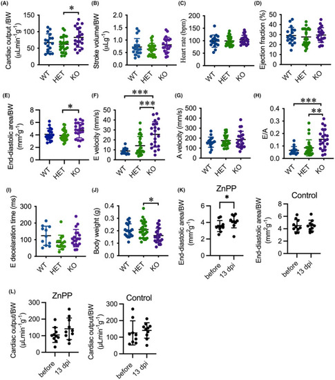
Deletion of hmox1a provokes cardiac output and hypertrophy in adults. (A–D) Echocardiography analysis depicting elevated cardiac output in KO(52del) zebrafish compared to their HET sibling (A) with a tendency toward increase in stroke volume (B) and no significant change in heart rate (C) and ejection fraction (D). (E) KO(52del) zebrafish exhibits enlarged end‐diastolic area compared to HET siblings. (F–H) PWD analysis indicating increased peak E wave velocity (F) and E/A ratio (H) without significant change on Peak A wave velocity (G). (I) Deceleration times of E wave from maximum velocity to baseline display no significant difference between the three genotypic groups. (J) KO(52del) zebrafish are lean in comparison with their HET siblings. (K) ZnPPIX treatment leads to enlarged end‐diastolic area in WT zebrafish at 13 dpi. Zebrafish treated with saline display no change at 13 dpi. (L) ZnPPIX treatment induces a tendency toward increase in cardiac output in WT zebrafish at 13 dpi. Zebrafish treated with saline display no change at 13 dpi. (A–J) WT n = 18; HET(52del) n = 25; KO(52del) n = 21. K, L ZnPPIX n = 10; Control n = 10. Data are presented as mean ± SD. One‐way ANOVA with Tukey adjustment for multiple comparisons in A–J. Two‐sample t‐test in K, L. *p < 0.05, **p < 0.01, ***p < 0.001.
|
