
Fig. 2
- ID
- ZDB-FIG-231110-39
- Publication
- Hagedorn et al., 2023 - Transcription factor induction of vascular blood stem cell niches in vivo
- Other Figures
- All Figure Page
- Back to All Figure Page
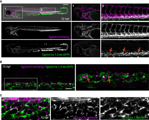
Niche endothelial-enriched mrc1a 1.3kb:GFP expression (A) Images show a double transgenic embryo carrying the pan-endothelial marker kdrl:mCherry (magenta) and the mrc1a 1.3kb:GFP transgene (green). Magnifications of boxed areas are shown on the right. The highest levels of vascular GFP expression are observed in CHT ECs (red arrows), whereas lower levels of expression are observed in the anterior head region, although some of these cells do not express the kdrl:mCherry transgene. (B) Images show runx1:mCherry+ HSPCs (magenta) directly interacting with mrc1a 1.3kb:GFP+ ECs within the CHT niche (red arrows). Panel on right shows magnification of boxed area. (C) cxcl12a:dsRed2+ stromal cells (magenta) are closely associated with mrc1a 1.3kb:GFP+ ECs in the CHT. Scale bars in (B) and (C) represent 100 μm. |
Reprinted from Developmental Cell, 58(12), Hagedorn, E.J., Perlin, J.R., Freeman, R.J., Wattrus, S.J., Han, T., Mao, C., Kim, J.W., Fernández-Maestre, I., Daily, M.L., D'Amato, C., Fairchild, M.J., Riquelme, R., Li, B., Ragoonanan, D.A.V.E., Enkhbayar, K., Henault, E.L., Wang, H.G., Redfield, S.E., Collins, S.H., Lichtig, A., Yang, S., Zhou, Y., Kunar, B., Gomez-Salinero, J.M., Dinh, T.T., Pan, J., Holler, K., Feldman, H.A., Butcher, E.C., van Oudenaarden, A., Rafii, S., Junker, J.P., Zon, L.I., Transcription factor induction of vascular blood stem cell niches in vivo, 1037-1051.e4, Copyright (2023) with permission from Elsevier. Full text @ Dev. Cell