FIGURE

Figure 4—figure supplement 1.

ID
ZDB-FIG-231101-5
Publication
Bellegarda et al., 2023 - The Reissner fiber under tension in vivo shows dynamic interaction with ciliated cells contacting the cerebrospinal fluid
Other Figures
All Figure Page
Back to All Figure Page
Figure 4—figure supplement 1.

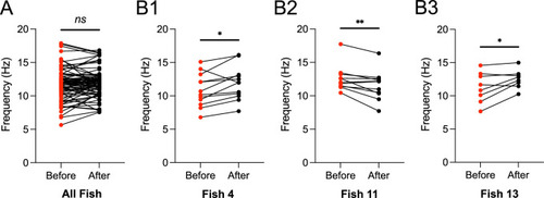
Changes in the main ciliary beating frequency varied from fish to fish in response to RF photoablation.

(A) The main beating frequencies of dorsal and ventral motile cilia after RF photoablation (mean ± standard deviation provided hereafter: 11.9 Hz ± 2.0 Hz) compared to those before RF photoablation (11.7 Hz ± 2.4 Hz), illustrating that cilia main beating frequency was on average not significantly affected by RF photoablation (N=76 cilia from 9 fish; paired two-tailed t-test: p > 0.3). (B1–B3) Select examples from (A), depicting fish whose response to RF photoablation significantly changed the main ciliary beating frequencies but in different ways. B1 and B3 illustrate an increase in main ciliary beating frequency after RF photoablation (B1: mean ± standard deviation provided hereafter: 11.9 Hz ± 2.5 Hz; B3: 12.5 Hz ± 1.4 Hz) from that before RF photoablation (B1: 10.9 Hz ± 2.5 Hz; N=12 cilia; paired two-tailed t-test: p < 0.05; B3: 11.3 Hz ± 2.3 Hz; N=8 cilia; paired two-tailed t-test: p < 0.05). B2 illustrates a decrease in main ciliary beating frequency after RF photoablation (11.6 Hz ± 2.2 Hz) from that before RF photoablation (12.6 Hz ± 1.9 Hz; N=11 cilia; paired two-tailed t-test: p < 0.01). * p < 0.05, ** p < 0.01.

Expression Data

Expression Detail
Antibody Labeling
Phenotype Data

Phenotype Detail
Acknowledgments
This image is the copyrighted work of the attributed author or publisher, and ZFIN has permission only to display this image to its users. Additional permissions should be obtained from the applicable author or publisher of the image. Full text @ Elife