|
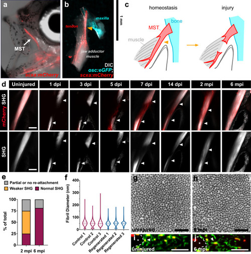
The adult zebrafish can fully regenerate after acute injury. a Epifluorescence image of an adult scxa:mCherry zebrafish with a brightfield overlay to demonstrate the position of the maxillary superficial tendon (MST) (denoted by white arrow). b Epifluorescence image of a normal uninjured MST musculoskeletal circuit (i.e. maxilla, MST, jaw adductor muscle (A0)) dissected from an osc:eGFP;scxa:mCherry adult zebrafish with a DIC overlay. The Orange arrowhead denotes where the injury is made. c Graphical schematic of the MST before and after injury. d 2-photon images of the MST at various time points after injury in scxa:mCherry zebrafish with SHG signal both overlaid and shown separately. White arrowheads denote the severed tendon ends. Scale bar, 100 µm. e Stacked bar graph showing the breakdown of zebrafish exhibiting partial or no reattachment, weaker SHG signal, or fully restored SHG signal at 2- and 6-months post-injury (mpi) (2 mpi: N = 12; 6 mpi: N = 16). f Violin plot illustrating the collagen fibril diameter distribution in 3 age-matched individuals uninjured and regenerated MSTs at 6 mpi. The means of uninjured controls 1, 2, and 3 were 53.33, 50.24, and 51.35 nm, respectively. Means of injured MSTs 1, 2, and 3 were 50.62, 50.31, and 49.41 nm respectively. Quartile values are shown with a dotted line and the median is shown with a dashed line. g, h Representative 50,000x TEM micrographs from age-matched uninjured (g) and regenerated tendons at 6 mpi (h). Scale bar, 500 nm. i, j Representative images of a cross-sectional re-slice view of 2-photon z-stacks from anti-mCherry stained (shown in green) scxa:mCherry MSTs from age-matched control uninjured (i) and regenerated MSTs at 6 mpi (j). White dotted lines outline muscle boundaries and asterisks mark muscles. Scale bar, 10 µm.
|