|
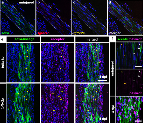
TGF-β signaling is active in tenocytes during regeneration. a–d Multiplexed RNA-scope in situ hybridization of scxa (green, a), tgfbr1b (red, b), and tgfbr2a (yellow, c) in the uninjured tendon. The merged image of all three is shown in (d). Scale bar, 50 µm. e Confocal images of anti-GFP immunofluorescence to detect CFP+ and/or YFP+ scxa-lineage cells (in green) combined with RNAscope in situ hybridization of either tgfbr1b (top panels) or tgfbr2a (bottom panels) in magenta at 4 days post-injury (dpi). Yellow arrowheads denote examples of co-positive cells. Scale bar, 50 µm. f Double immunostaining of anti-GFP immunofluorescence to detect CFP+ and/or YFP+ scxa-lineage cells (in green) with p-Smad3 staining (in magenta) in uninjured and regenerating tendons at 4 dpi. The middle panel shows the p-Smad3 staining alone in the uninjured tendon split from the merged image in the top panel. Orange and yellow arrowheads denote weakly p-Smad3+ nuclei in unlabeled and labeled tenocytes from lineage tracing, respectively. Scale bars, 25 µm.
|