IMAGE

Fig. 1

ID
ZDB-IMAGE-231002-83
Source
Figures for Tsai et al., 2023
Image
Figure Caption

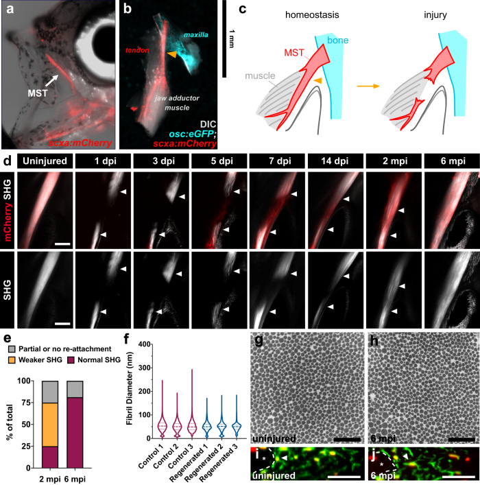
Fig. 1 The adult zebrafish can fully regenerate after acute injury.

a Epifluorescence image of an adult scxa:mCherry zebrafish with a brightfield overlay to demonstrate the position of the maxillary superficial tendon (MST) (denoted by white arrow). b Epifluorescence image of a normal uninjured MST musculoskeletal circuit (i.e. maxilla, MST, jaw adductor muscle (A0)) dissected from an osc:eGFP;scxa:mCherry adult zebrafish with a DIC overlay. The Orange arrowhead denotes where the injury is made. c Graphical schematic of the MST before and after injury. d 2-photon images of the MST at various time points after injury in scxa:mCherry zebrafish with SHG signal both overlaid and shown separately. White arrowheads denote the severed tendon ends. Scale bar, 100 µm. e Stacked bar graph showing the breakdown of zebrafish exhibiting partial or no reattachment, weaker SHG signal, or fully restored SHG signal at 2- and 6-months post-injury (mpi) (2 mpi: N = 12; 6 mpi: N = 16). f Violin plot illustrating the collagen fibril diameter distribution in 3 age-matched individuals uninjured and regenerated MSTs at 6 mpi. The means of uninjured controls 1, 2, and 3 were 53.33, 50.24, and 51.35 nm, respectively. Means of injured MSTs 1, 2, and 3 were 50.62, 50.31, and 49.41 nm respectively. Quartile values are shown with a dotted line and the median is shown with a dashed line. g, h Representative 50,000x TEM micrographs from age-matched uninjured (g) and regenerated tendons at 6 mpi (h). Scale bar, 500 nm. i, j Representative images of a cross-sectional re-slice view of 2-photon z-stacks from anti-mCherry stained (shown in green) scxa:mCherry MSTs from age-matched control uninjured (i) and regenerated MSTs at 6 mpi (j). White dotted lines outline muscle boundaries and asterisks mark muscles. Scale bar, 10 µm.

Acknowledgments
This image is the copyrighted work of the attributed author or publisher, and ZFIN has permission only to display this image to its users. Additional permissions should be obtained from the applicable author or publisher of the image. Full text @ NPJ Regen Med