|
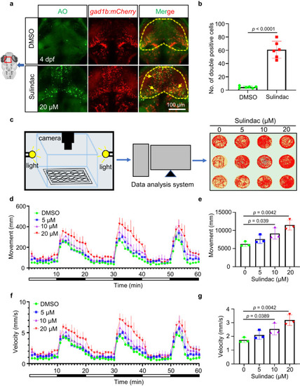
Sulindac selectively induced apoptosis of GABAergic neurons and altered motor behaviour in zebrafish larvae. a Most of the AO+ cells (green signals) were colocalized with mCherry+ GABAergic neurons (red signals) in the midbrains of Tg(gad1b:mCherry) embryos after sulindac treatment indicated by yellow arrows. b Quantification of the double-positive cells (yellow signals) in the midbrains of embryos. The values represent the means ± SDs (n = 6 larvae for per group, each dot denotes one larva). Statistics calculated by unpaired two-tailed Student’s t test. Source data are provided as a Source Data file. Apoptosis of GABAergic neurons in the midbrain is indicated by yellow arrows. c A system of behavioural experiments for recording larval motion trials in 60 min, and three representative photographs are shown for each group. d, f Swimming distance and velocity behavioural data were binned into 1-min intervals for analysis. The total movement (e) and average velocity (g) of zebrafish larvae exposed to sulindac at 5 dpf were examined during the light–dark photoperiod stimulation test (60 min). All data are presented as the means ± SDs (n = 3 independent biological replicates, eight larvae per replicate). Statistics calculated by unpaired two-tailed Student’s t test. Source data are provided as a Source Data file.
|