
Fig. 4
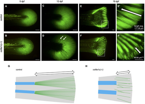
Fig. 4. Decreased density of actinotrichia and disorganized placement during the growth of col9a1c(-/-) fish. In vivo visualization of actinotrichia with actinodin1-GFP (Kuroda et al., 2018). (A, B) 5 dpf. (C, D) 12 dpf. (E, F) 19 dpf. A, C and E are control. B, D, and F are col9a1c(-/-). White arrows in D indicate abnormally thicker actinotrichia. (E′, F′) Magnified images of white dashed box in E and F, respectively. The number in each image represents the average of actinotrichia length (N = 10 each from control and col9a1c(-/-); t-test, ∗∗∗p < 0.001). The white double-headed arrows indicate the tip of the fin, an area where the fin-ray has not yet formed. This part is physically supported by the actinotrichia. (G, H) Schematic illustration at the fin tip. The fin is shown in grey. The blue boxes and green lines indicate fin-rays and actinotrichia, respectively. White double-headed arrows indicate the actinotrichia region. Scale bars: 100 μm. |
| Gene: | |
|---|---|
| Fish: | |
| Anatomical Term: | |
| Stage Range: | Day 5 to Days 14-20 |
| Fish: | |
|---|---|
| Observed In: | |
| Stage Range: | Day 5 to Days 14-20 |
Reprinted from Developmental Biology, 481, Nakagawa, H., Kuroda, J., Aramaki, T., Kondo, S., Mechanical role of actinotrichia in shaping the caudal fin of zebrafish, 52-63, Copyright (2021) with permission from Elsevier. Full text @ Dev. Biol.