
Fig. 3
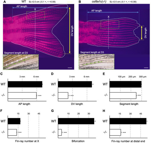
Fig. 3. Disturbed fin-ray configuration in adult col9a1c(-/-) zebrafish. (A, B) Representative fin images of WT and col9a1c(-/-) fish. Ten fishes were used for this measurement from each group and the standard length was adjusted to 2.5 mm (-0.1 <, < +0.08). Fin-rays were stained with Alizarin Red. AP length was measured as the distance (blue double-headed arrow) from the root of fin-rays to their most elongated tip (blue dot lines). DV length was measured as the distance (yellow double-headed arrow) between the most elongated tip of the fin-rays in each region of dorsal and ventral (yellow dot lines). The white dotted lines A and B indicate the position of X where the fin-ray outside D1 or V1 stops extending. The five segments comprising the third fin-ray from the dorsal side (D3), around the position X, were used to measure segment length (green dashed box). (C–G) The results of the analysis; AP length, DV length, segment length, the number of fin-rays at X, bifurcation, and the number of fin-rays at the distal end, respectively. Scale bars: 500 μm. (t-test, ∗∗∗p < 0.001). |
| Fish: | |
|---|---|
| Observed In: | |
| Stage: | Adult |
Reprinted from Developmental Biology, 481, Nakagawa, H., Kuroda, J., Aramaki, T., Kondo, S., Mechanical role of actinotrichia in shaping the caudal fin of zebrafish, 52-63, Copyright (2021) with permission from Elsevier. Full text @ Dev. Biol.